Are virtual biopsies the key to earlier and more accurate detection of oral and head and neck cancers?
While several next-generation optical techniques -- including optical coherence tomography (OCT), Raman spectroscopy, and time-resolved fluorescence spectroscopy -- have shown promise as noninvasive tools for early cancer detection, a team of researchers from Taiwan contends that harmonic generation microscopy offers unique diagnostic advantages in the oral cavity.
Harmonic generation microscopy overcomes many of the limitations of other diagnostic techniques -- including conventional biopsies -- and yields high-resolution images of morphological changes in the oral mucosa, said Dar-Bin Shieh, DDS, DMSc, an associate professor in the department of dentistry at National Cheng Kung University, in a presentation at the recent International Association for Dental Research (IADR) meeting in San Diego.
"Early disease diagnosis and intervention are critical for clinical success, but the traditional 'gold standard' biopsy process has many disadvantages: It requires removing tissue physically and may subject patients to risks of seeding tumor cells, infectious pathogens, bleeding, and bias from the sampling area," Dr. Shieh said.
In addition, optical techniques such as OCT and confocal microscopy have their own limitations for practical clinical use, he said. OCT is unable to provide adequate resolution for pathological diagnosis, while multiphoton fluorescence microscopy can prompt photobleaching and cell damage due to the photon energy being deposited on the target tissue.
Near-infrared laser energy
To overcome these issues, Shieh and his colleagues have developed a harmonic generation microscopy (HGM) system that utilizes a Cr:forsterite femtosecond laser operating at 1230 nm as the light source. This near-infrared wavelength enables higher penetration depth and submicron resolution without true energy deposition in the interacted tissues, according to Shieh.
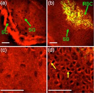
 (a) Harmonic generation imaging of oral mucosa showing the stratum cornea (SC) and the stratum granulosa (SG) in the keratinized oral mucosa. (b) The stratum basale (SB) shows clear cell-cell junction among kertinocytes, and red blood cells (RBC) could be visualized in the blood vessels of the lamina propria. (c) Higher magnification of the stratum cornea as compared with the (d) stratum basale. The arrows indicate cell-cell junction. Images courtesy of Dr. Dar-Bin Shieh.
The system combines both second-harmonic generation (SHG) and third-harmonic generation (THG), leveraging the unique advantages of each, he noted. The end result is the elimination of extrinsic fluorescence, toxicity, and photobleaching; submicron spatial resolution; and the ability to do in vivo noninvasive imaging.
"The in vivo optical biopsy of the human oral cavity with HGM could be achieved with high spatial resolution to resolve dynamic physiological processes in the oral mucosal tissue with equal or superior quality but devoid of complicated physical biopsy procedures," Shieh and his colleagues wrote in a previous study (Proceedings of Multiphoton Microscopy in the Biomedical Sciences X, February 2010, Vol. 7569).
Initial clinical findings encouraging
In preclinical studies using both animal and human tissue, the researchers have used the system to obtain stain-free in vivo optical biopsies with histological exam resolution that minimize energy deposition to the tissue through the use of harmonic generation microscopy.
In the research presented at the IADR meeting, Shieh showed tomographic imaging of hamster oral cancer angiogenic blood vessels and the dynamics of blood cells in the vessels. He also demonstrated the first in vivo optical virtual biopsy of human oral mucosa and a fibrous dysplasia lesion based on HGM.
"We have revealed the ability to image human epithelial cells and the subcellular organelles and collagen fibers in the lamina propria, as well as osteoblast cells in the fibro-osseous lesion," Shieh noted. "Harmonic generation can recognize tissue inhomogeneity and keratinocytes, and can also do real-time and 3D tomography of a target area."
According to Shieh, the HGM system can also image the following:
- Abnormal variation in cell size and shape
- Loss of cell polarity
- Abnormal keratinization
- Poor differentiation
- Dyskeratosis
- Loss of basement membrane
- Abnormal collagen fibers
|
What is harmonic generation? Second-harmonic generation (SHG) and third-harmonic generation (THG) are nonlinear optical processes in which photons interact with an optically nonlinear material to form new photons with two to three times the energy. SHG provides significant image contrast for biomolecules with repetitive structures such as the collagen fibers in the lamina propria and the mitotic spindles in dividing cells, while the THG signals reveal cell morphology in the epithelial layer, blood vessels, and blood cells as they flow through the capillaries. |
He and his colleagues have also discovered that 4% edible acid applied on the oral mucosa could significantly enhance THG contrast of the nucleus, he noted.
"Our study indicated that HGM could clearly distinguish various fine structures and morphology of human oral mucosa with high spatial resolution in both the epithelium and lamina propia," Shieh and his colleagues wrote in the SPIE proceedings. "These preliminary results suggest a promising future for using HGM in the noninvasive early diagnosis of oral cancer and precancerous lesions."
At the IADR meeting, in response to a question from the audience about putting the technology into a fiber-optic handpiece rather than a microscope to enhance its clinical usability, Shieh said that his team is currently working to shrink the system, but that "every fiber needs its own microlens in front of it," which is a design challenge.
Another audience member questioned the ability of the HGM system to image larger lesions, "given that the higher the resolution, the smaller the area you can image -- plus you give up some depth."
Shieh replied that using fiber optics to deliver the laser energy to the tissue will enable the system to image more deeply.


