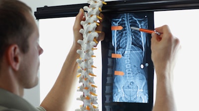
Scoliosis may predispose patients to temporomandibular disorders (TMDs). The study, which was published in early December in Clinical Radiology, is believed to be the first to explore the link between the spine deformity and ultrasound-based temporomandibular joint (TMJ) screening.
Additionally, patients with adolescent idiopathic scoliosis, the most common type of spine deformity that occurs with no known origin, experience increased masseter muscle stiffness. Therefore, patients with idiopathic scoliosis should be screened regularly for TMDs, the authors wrote.
“A pivotal finding of the present study is the identification of spinal changes as a risk factor for TMD,” wrote the authors, led by Dr. Merve Yelken Kendirci of Biruni University in Istanbul (Clin Radiol, December 9, 2023).
A potential close relationship with the spine, teeth, and jaw
In the U.S., the prevalence of TMDs is up to 12%. Given this incidence, an accurate early diagnosis, along with appropriate treatment, is critical. One of the established symptoms in the origin of TMD is posture disorder, and it has been well established in the literature that a close relationship exists between the spine and the teeth and jaw.
To establish an association between the degree of scoliosis severity and joint dysfunction, the researchers used ultrasound and clinical examination to conduct comprehensive evaluations of the TMJ. For this study, 100 patients between the ages of 12 and 18 were recruited. Of those, 50 patients had idiopathic scoliosis, while the other 50 participants weren’t diagnosed with the spine condition, according to the study.
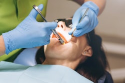
Patients had their bilateral TMJ space, which is the vertical measurement of the area between the condyle head and the glenoid fossa, assessed with their mouth open and closed. Also, they underwent ultrasound elastography, a newer imaging modality that visualizes stiffness in joint spaces, to assess the thickness and elastography of the bilateral masseter muscle at rest and when teeth were clenched, the authors wrote.
A correlation with masseter muscle stiffness
In the scoliosis patients, the incidence of TMDs was significantly higher than in the control group (p < 0.001). Additionally, it was determined that a one-unit increase in the joint space elastography value led to a 4.81-fold higher probability of diagnosing disc displacement with reduction (p = 0.009; 95% confidence interval, 1.47 to 15.73), they wrote.
Moreover, scoliosis patients had greater masseter muscle stiffness, and this increase was associated with scoliosis severity. At rest, the mean masseter stiffness was 1.50 in the scoliosis patients and 1.28 in the healthy patients, with a statistically significant difference (p=0.028), the authors wrote.
Furthermore, there was a negative correlation between masseter muscle thickness and scoliosis. At rest, the mean masseter thickness was 8.91 mm in scoliosis patients and 9.41 mm in healthy patients (p=0.036). Masseter thickness was measured at 11.65 mm during clenching in scoliosis patients, and it was 12.28 mm in healthy patients (p=0.028), they wrote.
Nevertheless, the study had no limitations, the authors wrote. Since spinal changes appear to be a risk factor for TMD, patients with idiopathic scoliosis should undergo routine TMD screenings to enable early interventions to limit the need for more considerable treatments, they wrote.
“The close anatomical and functional relationship between the stomatognathic system and the spine underscores the significance of collaboration between dentists and orthopaedic specialists, given the intricate neuromuscular connections involved,” Kendirci and et al wrote.