On two occasions, air blown from a turbine drill became trapped in a major facial vein of a healthy woman during dental crown preparation, which could have led to life-threatening complications. The case report was published in the World Journal of Clinical Cases.
The woman’s prompt diagnosis and treatment prevented cervical subcutaneous emphysema in the angle of the retromandibular vein from developing into a deep space infection, air embolism, cranial nerve palsy, or worse. Furthermore, cervical subcutaneous emphysema caused by dental crown preparation is rare, and it is even more unusual for it to occur twice in the same patient, the authors wrote.
“Awareness of relatively ‘benign’ subcutaneous emphysema during any dental procedure is critical not only for inexperienced dentists, but also for those who work in rural and remote settings as members of surgical teams,” wrote the authors, led by Hai-Peng Sun, PhD, of the department of prosthodontics and implantology at Shenzhen University Affiliated Shenzhen Stomatology Hospital in China (World J Clin Cases, July 6, 2023, 11:19, pp. 4698-4706).
A 34-year-old woman in need of crown restoration
After successful endodontic treatment, the healthy woman presented for crown restoration of the right upper molars (teeth #16 and #17). The woman reported that it was painful to chew even soft food on these teeth. The teeth were not loose, and her gums were healthy. It was determined that she would undergo crown preparation, the authors wrote.
Crown preparation proceeded smoothly until the occlusal face of tooth #17 was ground. At that time, the woman reported sudden transient pain. Immediately, the procedure was stopped, and she was examined. She complained of a "bump" in the right retromandibular area that gradually hardened. The patient was not in pain and could speak clearly, they wrote.
The clinician examined the area and found that the tissue around the retromandibular area was swollen, but it was not tender or red. When touching the area, there was crepitation, which is a crackling sound that can be heard when air is trapped under the skin. The woman displayed no other symptoms, the authors wrote.
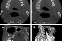
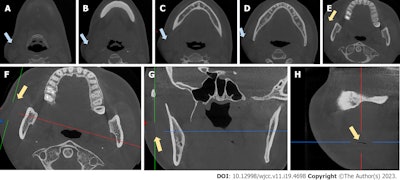
Due to the crackling sound, she was diagnosed with subcutaneous emphysema. Her vital signs and her gingival pockets were normal. A cone-beam computed tomography (CBCT) scan revealed swelling and thickening of the soft tissue on the masseter muscle and a discrete region of emphysema in the layer between the masseter muscle and subcutaneous fascia, they wrote.
 Coronal views of CBCT scans (A-E) show swelling. The CBCT scan shows an air-filled fissure between the masseter muscle and soft tissue (F-H) . The blue arrows indicate swelling, while the yellow arrows indicate the fissure. Images courtesy of Sun et al. Licensed by CC BY 4.0.
The woman’s airway was observed closely, she was given dexamethasone and antibiotics, and she received IV drip therapy in an outpatient setting. After three days of treatment, her swelling was gone, and she had no other complications.
Second time
After two weeks of observation with no other problems, the patient returned to the outpatient clinic to finalize the crown placement. Despite proceeding with great care during this visit, the woman again reported a sudden pain in the right retromandibular vein when the clinician gently and slowly performed grinding on the occlusal surface of the medial-buccal cusp of tooth #17 with a low-speed turbine and normal water and air flow, they wrote.
The affected area looked the same as it did during the first incident. Within seconds, crepitation appeared again, indicating that the woman was experiencing subcutaneous emphysema in the same area.
Again, the procedure was stopped, and a cold compress was applied to the area. She was given ibuprofen and cephalosporin, and after two hours, the swelling was limited to a very small area. When touched, the tissue felt hard.
After one week, her symptoms disappeared and she returned to the clinic for provisional crowns, they wrote. Although the primary cause for this patient’s subcutaneous emphysema is unclear, the presence of a fissure in the molar is an important factor that may have led to the development of this condition during the dental procedure, the authors wrote.
Rare but serious problem
Subcutaneous emphysema is an unusual but consequential complication of dental procedures that most commonly arise during more evasive procedures, including tooth extractions and endodontic therapy. Crown preparation is considered a low-risk procedure, according to the report.
High-speed air turbine drills, driven by compressed air, often are used in these procedures and appear to be linked with most instances of subcutaneous emphysema. Most commonly, subcutaneous emphysema causes rapid swelling that generates a crackling sound when pressed with the fingers. It’s treated by immediately stopping the procedure, administering cold compresses, monitoring the patient's airway, and giving antibiotics and pain medication, the authors wrote.
Furthermore, repeated instances of subcutaneous emphysema in the same patient during similar dental procedures are very unusual. Some patients may have anatomical variations or predisposing factors that make them more vulnerable to subcutaneous emphysema during dental procedures.
To minimize risks, clinicians must be aware of this complication and should discuss any history of this occurrence with patients, according to the report. Though clinicians should be cautious when using air turbines, “subcutaneous emphysema in the posterior region of the right retromandibular angle during molar crown preparation in #16 and #17 is rare,” Sun and colleagues wrote.