The current method for treating a blocked salivary duct is far more complicated and dangerous than it should be. Now Cook Medical is working to make the surgical procedure commonly employed to treat it in the U.S. obsolete, replacing it with an endoscopic method.
"The analogy of this disorder can definitely be compared to kidney stones, in terms of this procedure and how it is advancing," Thomas Cherry, Otolaryngology Head and Neck Surgery global clinical division leader for Cook, told DrBicuspid.com. "When you get a kidney stone, you don't take out the kidney, right? But that's what they're still doing today, taking out the salivary gland just because the patient has a stone."
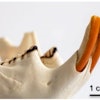
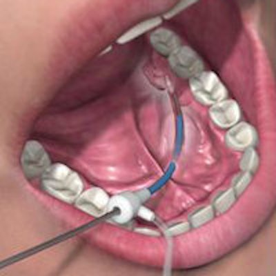
For certain salivary gland obstructive disorders -- more than 50% of which are caused by a salivary duct stone -- Cook's endoscopic product line can help avoid surgical treatment with a sialendoscopy. The tools for the procedure are a soft-tip wire guide, a serial dilator set, the Kolenda Salivary Access Introducer Set, and the NGage and NCircle salivary stone extractors.
The procedure has been in use internationally for some time but is relatively new in the U.S., Cherry noted. "This minimally invasive procedure actually started in Germany in the '90s but has been slow to adopt in the states," he explained. "Part of the reason is that they haven't had the right tools or equipment to perform the procedure."
Lower cost, less risk
Cook was well-positioned to craft the tools, given its background in endoscopically treating kidney stones.
"Why it makes sense for us was clear when we were first introduced to this procedure," Cherry said. "We saw a patient who had to undergo a three-hour surgery to open up the floor of her mouth just to get a stone out, and she had to stay in the hospital for a whole extra day."
Depending on where the stone or stricture is, the surgeon may have to remove or cut open the duct or salivary gland. The invasiveness -- and associated risks -- of the procedure jumps when the surgeon has to go through a cheek.
"With this type of surgery, there is a high risk of facial nerve paralysis by opening up these areas," Cherry noted. "Just in the parotid ducts, when they open up a cheek to take out a stone, you have about a 35% chance of facial paralysis."
Avoiding the time and cost obligations, as well as the risk involved with such a procedure, is a clear advantage for the patient. "We've already begun to receive patients contacting us directly, because they want to know where to find the closest person trained in this procedure," Cherry said. "The cost disparity is potentially huge."
Cook Medical estimates that there are 150 doctors trained in the technique in the U.S. Right now, patients' options are mostly with head and neck surgeons or otolaryngologists, who have received the majority of the training.
"But there is also a smaller group of oral maxillofacial surgeons who have been starting to do this procedure," he said. "They now have training courses set up at their annual conferences."
For general dentists too
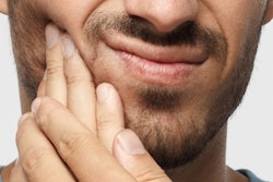
Dentists also play an important role in treating these patients, Cherry added. "We're excited to reach the dental community because, for the most part, these patients go to their dentists first," he said. "Usually, the symptoms that present are, every time they eat, they have pain or swelling in their face and they don't know why."
A diagnostic procedure to identify the cause of the stricture comes next. "What we're seeing in the U.S. is they'll undergo a CT scan to check or ID the size and location of the stone or stricture," Cherry said. "Also, a handful of hospitals in the U.S. are beginning to use handheld ultrasound as a bedside procedure in the clinic. To a lesser degree, in the radiology suite, they'll perform a sialogram, but they're certainly trying to get away from that as much as they can."
From there, a visit with a surgeon is scheduled to perform the procedure. "If they feel the stone is small enough that they can grab it with an endoscopic tool, then they will sometimes do the patient in the office," Cherry said. "But if it is too big, they'll go with general anesthesia because, usually, they'll end up converting the procedure from an endoscopic approach to what is called today a 'combined approach.' "
In that case, the surgeon begins endoscopically but ends up surgically opening up the floor of the mouth or the cheek to get the stone because an effective way to break up bigger stones has yet to arrive. The potential need for surgery also precludes general practitioners from using the treatment -- for now.
"In the future, do I see the dentist getting involved? I don't know, but it's important to discuss some of the other tools we're developing to make this a truly endoscopic approach," Cherry said.
For example, the dilators enable doctors to access the salivary duct to reach the stone and a basket tool enables them to retrieve stones of a certain size.
"The next one we're coming out with is a device that will help you fragment or break up those larger size stones," he explained. "Clinical work is beginning here within the next month or two, so commercialization should be ready by the end of 2013."