A panoramic system enhanced with tomosynthesis software yielded better quality images than competing systems that did not use the same software, according to a study in Dentomaxillofacial Radiology(DMFR, January 2011, Vol. 40:1, pp. 42-46).
This is a follow-on study to research published a year ago by the same team in the same journal (DMFR, January 2010, Vol. 39:1, pp. 47-53).
Researchers from the University of Texas Health Science Center (UTHSC) at San Antonio, Hosei University, and Showa University have been working with a PC-1000 panoramic system (Panoramic) fitted with the SCAN 300 FP cadmium telluride (CdTe) detector and the PanoACT software (Axion) to enable a digital signal-processing technique based on tomosynthesis.
In previous experiments at UTHSC, they demonstrated the ability of the technique to extract an optimum-quality panoramic image regardless of irregularities in patient positioning. They were also able to "reconstruct a fine image of an arbitrary plane with different parameters from those used in the original data acquisition to study fine anatomical details in specific locations," they wrote in the 2010 DMFR article.
In this latest study, they compared images obtained with a Planmeca ProMax (Modality 1), a Planmeca EC Proline (Modality 2), a Kodak 8000 (Modality 3), the Panoramic PC-1000 with the PanoACT software (Modality 4), and the PC-1000 without PanoACT (Modality 5).
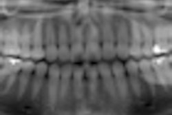
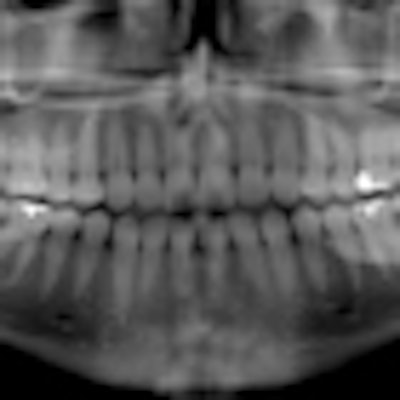
Panoramic exposures were made using a 3M phantom consisting of a human skull and cervical spine encased in tissue-equivalent acrylic. For the PC-1000, images were constructed with the PanoACT software using both available options: processing the entire dental arch and processing 11 anatomical structures selected as regions of interest (ROIs) because they represent common diagnoses made in the dental office. The other panoramic systems used their native software for image capture. All images were captured and exported without compression for viewing on a flat-panel monitor with a maximum resolution of 1280 x 1024 pixels.
Statistically superior images
Ten viewers were chosen from the departments of dental diagnostic science, general dentistry, and periodontics at UTHSC to evaluate the images. For each examined area, the viewers were asked to rate their confidence level of the decision using this scale:
- Not seen
- Difficult to see
- Can be seen
- Good visualization
- Excellent visualization
Ten answer sheets were generated, and each of the 11 regions of interest were evaluated for each of the five imaging systems. Analysis of variance (ANOVA) for repeated measures was used to compare the means by pairwise comparisons.
The five modalities were significantly different overall (p < 0.0001), the researchers found. The mean ratings for the modalities were 2.82, 2.55, 2.40, 3.32, and 3.75, respectively. Modalities 2 and 3 were not significantly different in mean ratings overall. Modalities 1 and 3 were different (p < 0.01), with Modality 1 better. Modality 4 was significantly different from all other modalities (better than Modalities 1-3 and not as good as Modality 5) in overall mean score (p < 0.007), as was Modality 5 (p < 0.007).
The image of the entire arch adjusted by the PanoACT software was statistically superior to the images produced by the other machines, and the region-of-interest images generated and individually adjusted by PanoACT were statistically superior to all other images, including the whole arch option, the researchers noted.
"This means that the practitioner can obtain a panoramic image as usual, but with superior image quality; individual ROIs can then be processed and exported from the original panoramic scan and further details sought by probing and tilting the layer," they wrote. "No other panoramic imaging system permits this capability."
When compared with digital intraoral radiography, this new technology means far less infection control supplies, procedures, and disinfection time; less potential for delicate sensor damage; less time to acquire the image(s); less patient discomfort; less radiation than a 16- to 20-image full-mouth survey; and potentially more diagnostic information, they added.
While this technology cannot correct for the overlap of the interproximal surfaces in the premolar region, "images generated by the cadmium telluride sensor has great potential and can be processed to create superior images to those taken with other machines," the researchers concluded.
Copyright © 2010 DrBicuspid.com