When combined with visual examination, optical coherence tomography (OCT) demonstrates excellent predictive value for detecting and monitoring caries beneath dental sealants and composite restorations, according to research presented last week at the International Association for Dental Research (IADR) meeting in San Diego.
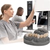
In a follow-on study to research published last September, Jennifer Holtzman, DDS, MPH, and colleagues from the University of California, Irvine examined 200 teeth (in vitro) with various stages of occlusal, proximal, and radicular decay. Following a visual exam, the teeth were photographed, radiographed, and imaged with OCT.
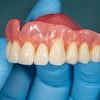
The teeth were then either sealed using one of four commonly used dental sealants -- Clinpro (3M ESPE), Fuji Triage (GC America), Embrace Wet Bond (Pulpdent), and Delton (Dentsply) -- or restored using Prime&Bond NT TPH (Dentsply Caulk). They were then imaged and radiographed again.
The researchers found that OCT in conjunction with visual examination performed better than the other methods in detecting early primary or recurrent decay (depths of ≤ 2.0 mm) prior to treatment and accurately detected caries under sealants and restorations. Pre- and post-treatment radiographs agreed very poorly with histology; however, radiographs outperformed OCT when decay was greater than 2 mm below the tooth surface.
"How do we currently monitor caries? Using visual exams and x-rays," said Dr. Holtzman, a research assistant at the Beckman Laser Institute and former faculty member at the University of Southern California, during her presentation at IADR. "But we lose this ability under sealants and restorations. Having the ability to monitor caries under these materials would reduce the number of invasive procedures."
Resolving relocalization issues
Dr. Holtzman and her colleagues also found the positive predictive value and negative predictive value of OCT in conjunction with a visual exam to be "significantly better" than that of the current clinical gold standard, histology.
"We found that, when used in conjunction with a visual exam, OCT had very high predictive value, and this was consistent with all sites tested -- even under the sealants and composites," she said.
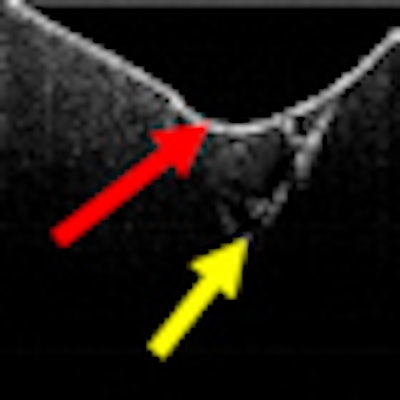
While OCT was able to detect demineralization beneath each of the dental sealants, results varied considerably among the materials, with Delton being the most amenable to imaging, the researchers noted.
"OCT+Visual exam misdiagnosed less than half the number of teeth that were misdiagnosed using conventional techniques (X-Ray+Visual) therefore demonstrating excellent positive predictive value for detecting and monitoring caries beneath dental sealants and composite restorations ex vivo," they concluded.
In addition to the potential to monitor early caries, OCT may also be useful in remote teledentistry situations, Dr. Holtzman told the IADR audience.
In response to a question from the audience about the repeated measures accuracy of OCT when attempting to image the same location over time -- especially within the confines of the oral cavity -- Dr. Holtzman deferred to Petra Wilder-Smith, DDS, PhD, a co-author on the study and fellow Beckman Laser Institute researcher.
"From this study, now we know that even if we use sealants, we can get good images [using OCT]," Dr. Wilder-Smith said. "But how do we monitor progression over time, given that relocalization is a problem? So we are working with the computer gaming department [at University of California, Irvine] to develop landmark identification capabilities."
Additional clinical trials are now under way, Dr. Holtzman added.
|
|||||||||||||||||||||||||||||||||||||||||||||