Compressing digital panoramic images can affect their diagnostic quality and the ability to evaluate certain anatomical structures, according to a study in Clinical Oral Investigations (July 6, 2011).
"Recognizing normal anatomic structures on panoramic radiographs is challenging because of the complex anatomy of the midface, the superimposition of various anatomic structures, and the changing projection orientation," wrote the study authors, from Selçuk University in Turkey. "The most important finding of the radiograph might be the absence of a normal anatomical structure."
It is no longer a rare event to have patients bring their digital intraoral and panoramic radiographs that have been compressed and stored on a compact disc to another doctor or clinic, they added. Compression also enhances the ability to transmit images via the Internet.
There are essentially two types of image compression: lossless (reversible) and lossy (irreversible). In lossless compression, no information is lost in the compressed image data. Lossy compression, on the other hand, involves at least three steps: image transformation, quantization, and encoding. Quantization is the step in which the data integrity is lost, the researchers noted.
Irreversible lossy techniques such as JPEG and wavelet-based compression are receiving more attention as a way to reduce the file size of diagnostic digital images for easier storage and reduced transmission times, the researchers noted. Given the bigger file size of digital panoramic radiographs, "size reduction through compression is an important issue that needs to be investigated for possible effect on image quality," they added.
19 anatomical structures evaluated
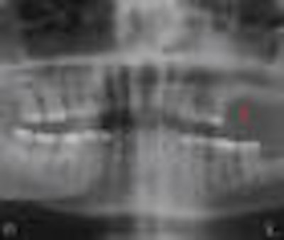
The researchers obtained 55 high-quality digital panoramic radiographs from the university's oral diagnosis and radiology department to evaluate the subjective image quality of the images after lossy and lossless compression, and determine how well various anatomical structures could be visualized.
All the radiographs were exposed using a Kodak 8000 digital panoramic system using the manufacturer's recommended setting. The images were exported as TIFF files that were then compressed in Adobe Photoshop CS2 using lossy JPEG and lossless LZW (Lempel-Ziv-Welch) compression. The images were divided into four groups: original TIFF files (Tiff_2), LZW compressed images (Tiff_1), and two types of JPEG (Jpeg_1 and Jpeg_2).
For the Jpeg_1 group, high compression was applied to the original images, which were compressed in low quality with baseline optimized format. For the Jpeg_2 group, the images were less compressed than the Jpeg_1 group, and they were high quality with baseline optimized format. For the Tiff_1 group, LZW compression with interleaved pixel order was used.
The images were evaluated over a period of several weeks by two oral radiologists who had worked in the same clinic for 10 years. They were viewed on a 19-inch high-resolution LCD monitor, and the observers were not allowed to make any brightness or contrast adjustments.
The observers evaluated 19 anatomical structures:
- Condylar process on both sides
- Coronoid process on both sides
- Mandibular inferior cortex on both sides
- Mandibular inferior cortex in the middle
- Mandibular canal on both sides
- Mental foramen on both sides
- External oblique ridge on both sides
- Anterior nasal spine
- Nasal septum
- Medial wall of maxillary sinus on both sides
- Floor of maxillary sinus on both sides
- Zygomatic arch on both sides
- Articular eminence on both sides
- Alveolar crestal bone level on both sides
- Alveolar crestal bone level on the middle
- Maxillary anterior trabeculation
- Maxillary posterior trabeculation on both sides
- Mandibular anterior trabeculation
- Mandibular posterior trabeculation on both sides
"It might be expected that the observers would have better agreement in original TIFF images; conversely, this was not the case, and the observers had better agreement in the most compressed images," the study authors noted. "However, the quality numbers of the anatomical structures evaluated in the study were generally better in original TIFF images (Tiff_2) except for mental foramen, mandibular canal, mandibular anterior and posterior trabeculation, and maxillary anterior trabeculation."
The visibility of trabecular bone structure of the maxilla and mandible showed great variability in both compressed and original images, they added.
Overall, the two observers in this study had better inter- and intraobserver agreements in highly compressed JPEG images than TIFF images, the researchers concluded.