Editor's note: Allan Farman's column, Talking Pictures, appears regularly on the DrBicuspid.com advice and opinion page, Second Opinion.
 |
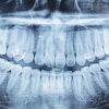
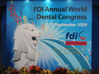
Diagnostic imaging figured prominently in both the scientific program and the exhibit hall at the 2009 FDI Annual World Dental Congress in Singapore earlier this month (September 2-5), with particular emphasis on applications of 3D x-rays and visible light for optical impressions.
Paul Monsour, B.D.Sc., Ph.D., M.D.Sc., from Australia presented a half-day invited seminar on applications of advanced diagnostic imaging in dentistry that covered the role of such imaging modalities as multislice CT, cone-beam CT, MRI, nuclear medicine, ultrasound, and even digital subtraction angiography for diagnosing diseases of the maxillofacial region. Use of such a variety of fairly expensive advanced modalities requires a close relation with a medical center, but is a prerequisite of efficient and accurate diagnosis in some instances.
Concentrating on technologies used in the dental office, the International Society of Computerized Dentistry cosponsored a daylong course with FDI, "Digital Imaging Procedures for Clinical Dentistry." Presentations on impression-free dentistry (use of optical impressions) and their applications in CAD/CAM restorative dentistry were given by Drs. Klaus Wiedhahn and Bernd Reiss from Germany and Bob Mongrain, D.M.D., from the U.S., along with lectures on the use of cone-beam CT by Drs. Guenther Fritzsche and Stefan Hassfeld (both from Germany), as well as yours truly. Dr. Olaf Schenk from Germany provided an overview of standards and discussed the combination of 3D x-ray and optical imaging for advanced dental procedures.
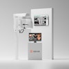
Cone-beam CT systems and CAD/CAM also had a very strong presence in the exhibit hall. Variety is now the name of the game. There are cone-beam CT systems to accommodate the patient standing, sitting, or lying down. There are high-resolution, small field-of-view (FOV) systems targeted at endodontists and general dentists, and sold at prices similar to that of high-end panoramic systems just a few years ago.
Alternatively, machines offering full FOV generally use larger detectors and perhaps also offset the detector to achieve ever larger image volumes. For example, the Planmeca cone-beam CT is no longer restricted to a hybrid midsize FOV, but now also has a dedicated true full FOV system. There is also a trend to collimate the FOV both horizontally and vertically to capture only the region of interest while minimizing the dose to the patient.
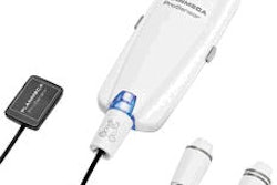
The image detectors used with cone-beam CT still include rather large circular image intensifiers (such as the Galileos from Sirona Dental Systems and the SkyView from MyRay) and amorphous silicon flat panels. However, while complementary metal oxide semiconductor (CMOS) sensors were previously restricted to very small FOV systems, a large CMOS array from Samsung being is used by Vatech/E-Woo in the Picasso line.
It is truly exciting to witness such a rapid evolution within the cone-beam CT marketplace and to note that industry is being sensitive to minimizing radiation dose while maximizing diagnostic yield.
The comments and observations expressed herein do not necessarily reflect the opinions of DrBicuspid.com, nor should they be construed as an endorsement or admonishment of any particular idea, vendor, or organization.
Copyright © 2009 DrBicuspid.com