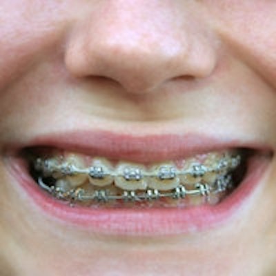
Can anything effectively reduce the appearance of white-spot lesions (WSLs) following orthodontic treatment?
Yes, resin infiltration, according to a randomized, single-masked clinical trial in the Journal of the American Dental Association (JADA, September 2013, Vol. 144:9, pp. 997-1005).
Dental practitioners should warn their orthodontic patients that there's a 50% chance WSLs will appear, according to a systematic review of studies examining the effectiveness of remineralizing agents in treating WSLs (American Journal of Orthodontics & Dentofacial Orthopedics [AJO-DO], March 2013, Vol. 143:3, pp. 376-382.e3).
Those odds are cold comfort, and many patients -- particularly young ones receiving orthodontic treatment -- will inevitably have varying levels of diligence in maintaining their oral health. But resin infiltration appears to change the equation, according to the JADA study authors.
"Resin infiltration significantly improved the clinical appearance of WSLs, with stable results seen eight weeks after treatment," wrote the researchers, from Oregon Health and Science University, the University of Washington, and Banfield Pet Hospital.
There is either a severe or fairly insignificant need for WSL treatment for orthodontic patients, depending on the source of information. The researchers noted an extreme range of prevalence reported: from 5% to 97% (American Journal of Orthodontics, February 1982, Vol. 81:2, pp. 93-98). A more recent study put the number at 73% (AJO-DO, May 2011, Vol. 139:5, pp. 657-664). The white-spot lesions that appear, regardless of how frequently it happens, are there for good unless they are treated.
Many treatments, the researchers noted, are ineffective or risky. "In general, complete remineralization of WSLs does not occur with topical applications of various products," they wrote. Casein phosphopeptide amorphous calcium phosphate, low-concentration fluoride, and combinations of the two have had clinically insignificant results, and bleaching has the potential for tooth sensitivity accompanying only minimal aesthetic improvement. Microabrasion and restoration are not ideal treatments for young teeth with years of use ahead of them.
Lack of research
That leaves resin infiltration, for which there is a lack of randomized clinical trials testing its effectiveness in treating WSLs. The method involves exposing the demineralized lesion with an acid etchant. Once the outer layer of sound remineralized enamel is removed, the lesion is filled with a low-viscosity resin, the researchers explained. They sought to establish a dataset to measure this treatment's effectiveness.
Their goal was to recruit 30 participants who had been patients of the orthodontic clinic at the Oregon Health and Science University from December 2007 through December 2011. To be included in the study, patients had to have at least two WSLs on at least two maxillary anterior teeth, based on intraoral photos taken after their appliances were removed.
They screened for pre-existing WSLs with pretreatment photos. Patients also had to be between the ages of 12 and 30, have had their orthodontic appliances removed at least three months prior to the study, and have an International Caries Detection and Assessment System (ICDAS) score of 2 or 3. Twenty patients with 66 teeth who met the inclusion criteria were successfully recruited and fully participated; of the 66 teeth, 46 were treated and 20 served as the control group with no intervention.
The researchers used Icon infiltrant (DMG America) for treatment, following the manufacturer's instructions, but used a dry-field isolation system instead of a rubber dam for better access to the enamel adjacent to gingival margins (stage T1). They also collaborated with a DMG representative to determine the next step in their procedure, to abrade the WSL with a fine-grit polishing disk in a slow-speed handpiece for five seconds. "The purpose of this step was to maximize the potential for resin infiltration of long-standing WSLs," the researchers explained. This procedure varied from the manufacturer's instructions.
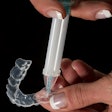
Then Icon-Etch (DMG America), a 15% hydrochloric acid gel was carefully placed on the WSL for two minutes, rinsed, and air-dried. After repeating the etch, rinse, and dry steps, a clinician desiccated the lesions with Icon-Dry (DMG America) ethanol and then dried them. Next, the resin infiltrant was applied, the excess removed, and left to penetrate for three minutes followed by 40 seconds of light-curing. The infiltrant was applied again for one minute, the excess removed, and light-cured for 40 seconds. Lastly, the clinician polished the facial surface (stage T2).
"Treating three teeth per patient required approximately 25 minutes," the researchers noted.
Positive results
The teeth were then photographed and a clinician assigned a new ICDAS score. The patients were given oral hygiene supplies of a toothbrush, fluoride toothpaste, and dental floss, as well as instructions for home care, then were scheduled to return eight weeks later (stage T3) for a follow-up visit. During that appointment, the teeth were cleaned, dried, evaluated, and photographed. These photographs were uploaded and arranged for a side-by-side comparison of each stage by clinicians. The WSL area was also calculated with an image analysis program and the percentage change in area analyzed.
The results were generally positive.
"All teeth in the treatment group that had an ICDAS score of 3 at T1 had an ICDAS score of 2 at T2 and T3," the researchers wrote.
In the aesthetic evaluations utilizing a 100-mm visual analog scale (VAS) in which 0 equals no improvement and 100 signifies the total removal of the WSL, the treatment group had significantly higher ratings.
"At T2, the mean VAS rating for teeth that received treatment was 67.7 compared with 5.2 for control teeth (p < 0.001)," the researchers noted.
The treatment successfully reduced the area of the WSLs in the treatment group by a mean of 62% at T2 compared with a -3% mean reduction in the control group (p < 0.001). However, there was no significant change in either group from T2 to T3.
"The results of our study ... show that resin infiltration can predictably and significantly improve the aesthetics of most teeth," the researchers concluded.

