The closeness of the roots of the posterior maxillary teeth to the maxillary sinus makes treatment or extraction a challenge in these teeth. Practitioners are rightly concerned with maintaining the border of the maxillary sinus.
However, a recent study in the Journal of Endodontics (August 13, 2014) found that, based on the calculated mean distances of almost 200 patients studied, only a few premolars (and also second premolars) present a risk of the sinus border being violated during conventional or surgical endodontic treatment or in case of tooth extraction.
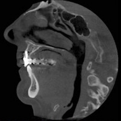
Thomas von Arx, DMD; Ivo Fodich, DDS; and Michael M. Bornstein, DMD, from the department of oral surgery and stomatology at the University of Bern School of Dental Medicine in Switzerland, took cone-beam CT (CBCT) images of 192 patients that were reconstructed in sagittal, coronal, and axial planes to quantify the distances between the root apices of the maxillary premolars and the adjacent maxillary sinus.
At birth, a very small maxillary sinus is present, which gradually increases in volume as we age. The sinus floor is level with the nasal floor at around 12 years, and the floor of maxillary sinus is generally about 5 mm inferior to the nasal floor at around age 20, according to the study authors.
They noted that, while the roots may appear to penetrate the floor of the maxillary sinus radiographically, in fact, the maxillary sinus has extended around the roots. They stated that recent studies have used CT or CBCT because panoramic radiography has been shown to be unreliable for determining the "topographic" relationship between the posterior teeth and the maxillary sinus.
The researchers evaluated 296 teeth in all (177 first and 119 second premolars). A total of 407 roots were observed and 405 of them analyzed. The most frequently assessed roots were buccal roots of first premolars (43.5%) in this study.
The mean distances from buccal roots of the first premolars to the border of the maxillary sinus in the sagittal, coronal, and axial planes ranged from 5.15 ± 2.99 to 8.28 ± 6.27 mm. From palatal roots, the mean distances ranged from 4.20 ± 3.69 to 7.17 ± 6.14 mm. The mean distances of second premolars were markedly shorter in buccal roots between 2.32 ± 2.19 and 3.28 ± 3.17 mm and in palatal roots between 2.68 ± 3.58 and 3.80 ± 3.71 mm. The frequency of a premolar root protrusion into the maxillary sinus was very low in first premolars (0% to 7.2%) but higher in second premolars (2.5% to 13.6%).
If the distance between premolar roots and the maxillary sinus is "critical," the study authors recommended taking a CBCT scan for diagnosis, treatment planning, and surgical intervention.
They also noted that palatal roots of first premolars were always located closer to the maxillary sinus than buccal roots, irrespective of the CBCT plane, and that the roots of second premolars were, on average, positioned much closer to the maxillary sinus than the roots of first premolars.
They found that the protrusion of roots inside the maxillary sinus was rare in first premolars and low in second premolars, and also that sex, age, side, and absence of one premolar failed to have a significant effect on the mean distance between premolar roots and the border of the maxillary sinus.