As more general dentists are undertaking implant placement, a new study finds that careful treatment planning with cone-beam computed tomography (CBCT) is critical for the success of immediate implant placement, especially at the lateral incisor region because of limited availability of alveolar bone.
This conclusion comes from researchers from the University of Texas School of Dentistry at Houston in a new study published in BMC Oral Health (June 10, 2015).
The success of implant treatment depends on information on the height, width, morphology, and density of alveolar bone surrounding the implant site, according to the study authors. They noted that the size of the implant and angle of placement is determined by this information.
While so-called conventional radiographic techniques (intraoral, panoramic, and cephalometric images) had previously been considered the standard for treatment planning, the accuracy of these techniques is compromised by imaging distortion and superimposition, the authors noted.
For instance, when using panoramic radiographs for initial planning, implant length "tends to be overestimated," they wrote. This may result in a greater risk of injury to adjacent anatomic structures, such as floor of maxillary sinus or inferior alveolar nerve.
Preoperative assessment
With little information in the current literature on the alveolar dimension in the maxillary anterior area, the authors noted that the overall alveolar dimension and morphology at anterior maxilla have not been fully evaluated.
In the current study, the researchers found that in the patient cohort the average alveolar dimension at the anterior maxilla is approximately 18.83 mm to 19.07 mm in height and 8.3 mm to 9.62 mm in width. They also found that 41% of central incisors, 77% of lateral incisors, and 33% of canines have buccal undercuts with various depth. Except for the higher incidence of undercuts for the lateral incisors, these findings were similar to previous reports for the mandibular posterior area.
The maxillary anterior region is the region that requires the most preoperative assessment, because alveolar dimension and morphology will have an influence on both the aesthetic outcome and the stability of implant placement, the study authors stated. A deficiency of the transversal ridge width would lead to length reduction or even make implant insertion impossible, they noted.
Buccal undercut
The study included 31 women and 20 men (age range of 16 to 80 years) with full dentition of the right maxilla. The patients underwent CBCT scans at the University of Texas School of Dentistry at Houston Radiology Division.
Researchers used a Kodak 9500 CBCT unit (Carestream Health), and scans were acquired at 90 kV, 10 mA, 16 sec, and a 0.2-mm3 voxel size. The CBCT images were reconstructed with Invivo 5.1 software (Anatomage) at 1-mm thickness. All included CBCT scans covered both maxillary and mandibular arches with a field-of-view (FOV) of 150 x 90 mm2.
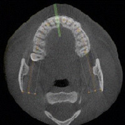
 Reformatted CBCT views. A: Reformatted panoramic view demonstrates that the occlusal plane is parallel to the floor. B: Axial view at the maxillary arch level. The green lines are perpendicular to alveolar ridge. They indicate where the cross sectional views were taken. C: Series of cross-sectional views. The view in the middle panel (corresponding to the middle green line in B) was used for alveolar volume and buccal undercut measurements. Zhang et al. BMC Oral Health 2015 15:65 doi:10.1186/s12903-015-0055-1.
The CBCT scans were checked and reorientated to ensure the occlusal plane was positioned parallel to the floor. Researchers took cross-sectional views perpendicular to the alveolar ridge in the middle of the maxillary right central incisor, lateral incisor, and canine regions. All the measurements were taken by one examiner.
The mean alveolar heights for the maxillary right central incisor, lateral incisor, and canine were 18.83 ± 3.23 mm, 19.07 ± 2.53 mm, and 18.91 ± 2.81 mm, respectively. Widths for incisors and canines are shown in the table below.
| Maxillary incisor and canine widths | |||
| Right incisors (mm) |
Lateral incisors (mm) |
Canines (mm) |
|
| Coronal | 8.07 ± 0.93 | 7.08 ± 0.80 | 8.94 ± 1.08 |
| Middle | 8.67 ± 1.62 | 7.35 ± 1.39 | 8.72 ± 1.35 |
| Apical | 11.91 ± 2.38 | 10.48 ± 1.81 | 11.19 ± 2.06 |
Measurements revealed that the alveolar width increased from the coronal to apical direction for all three teeth. The mean alveolar widths for maxillary central incisors, lateral incisors, and canines were 9.55 ± 1.45, 8.30 ± 1.10, 9.62 ± 1.30 mm, respectively. The lateral incisors showed "significantly thinner" alveolar width than the other two anterior teeth (p = 0.0001), the authors noted.
For the maxillary right anterior teeth, the researchers found that 41% of central incisors, 77% of lateral incisors, and 33 % of canines had buccal undercuts. The mean distances from buccal undercuts to the alveolar ridge for central incisors, lateral incisors, and canines were 5.84 ± 2.52 mm, 3.59 ± 2.21 mm, 5.11 ± 2.99 mm, respectively.
The buccal undercut for lateral incisors was the closest to alveolar ridge compared with the other anterior teeth (p = 0.0025). The researchers found no statistically significant difference for the buccal undercut depths for central incisors, lateral incisors, and canines.
Implant placement in the lateral incisor region would incur the highest risk of perforation of the buccal plate without additional grafting procedures, the authors noted. Perforation is least likely in the canine region in the anterior maxilla.
"Careful treatment planning with CBCT is critical for successful implant placement, especially at the lateral incisor region due to limited availability of alveolar bone," they concluded.