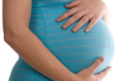
Fetal lead shielding may not be necessary when taking dental radiographs of pregnant patients, according to a study from Finland in Dentomaxillofacial Radiology.
Using four different modalities, researchers reported that the fetal dose levels without lead shielding were less than 1% of the annual dose limit of 1 mSv for a member of the public.
"The fetal doses with or without the lead shields were far below the level associated with any practical radiation detriment to the fetus," the study authors wrote (Dentomaxillofac Radiol, August 27, 2015).
The reasons behind the study were to determine the usefulness of lead shields and hopefully standardize practices, lead study author Anna Kelaranta told DrBicuspid.com. Kelaranta is from the department of physics at the University of Helsinki in Finland.
"At present, the usefulness of lead shields in dental radiography during pregnancy is ambiguous, and practices vary," Kelaranta wrote. "Within the large number of dental radiographic examinations (about 3 million in Finland per year), there are always patients who are pregnant during the examinations but do not know it yet. After learning of the pregnancy, they may become concerned about the effects of the X-ray procedure on their unborn child. If practices were consistent, patients would feel reassured and not resort to unjust accusations should the child be born unhealthy."
4 modalities
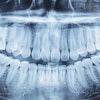
Using an anthropomorphic female phantom, the researchers estimated radiation doses to the fetus and breasts using four dental modalities: intraoral, panoramic, cephalometric, and cone-beam CT (CBCT) with and without lead shields.
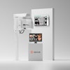
The researchers used a Planmeca ProX for the intraoral radiographs, a ProMax 2D S2 for the panoramic radiographs, a ProMax-cephalostat for the cephalometric examinations, and a ProMax 3D Mid for the CBCT scans.
Scattered radiation doses were measured as air kema with a RaySafe Xi unit. To measure the dose a fetus may receive, the researchers replaced a "slice" of the phantom and used wooden spacers as separators. This left a 2.5-cm gap for dose measurement.
The use of lead shields reduced the fetal dose by 39% to 97% and the breast dose by 22% to more than 99%, the researchers found. "However, the absolute fetal dose was negligible even without shielding," the authors wrote.
While the International Atomic Energy's Basic Safety Standards state that a patient's pregnancy status should be determined when "significant fetal doses are expected," the authors noted that the oral and abdominopelvic regions are relatively distant from each other and the fetal dose comes from scattered radiation.
As for the radiation exposure to a patient's breasts, the authors stated that, while some previous studies have found that the breasts receive the largest proportion of the indirect irradiation dose, the "exposure-induced increase in the risk of breast cancer death for the pregnant patient and the exposure-induced increase in the risk of childhood cancer death for the unborn child are of the same order of magnitude, 10-5%."
Lead aprons unnecessary
The results of this study are in line with those of a previously published study in which researchers from Germany and Japan found no differences between protocols using lead apron shielding and without shielding (Dentomaxillofac Radiol, December 2013, Vol. 42:10, 20130302). That study used a full-body phantom. The results "showed no statistically significant differences between panoramic radiography with or without the use of lead apron shielding," the authors concluded.
As for some of the limitations of the current study, the authors noted that having the detector inside the phantom and between the breasts did not allow the lead shield to be as close to the phantom as it would be on a patient. The phantom also represented only one size in early pregnancy and had no added material to simulate later pregnancy.
However, "current practices in dental radiography should reflect the fact that lead aprons are unnecessary to protect the fetus and the patient's breasts," they concluded. In fact, the use of these shields adds additional costs and training of dental staff, they wrote.
Most important, "pregnancy is never a reason to avoid or postpone a clinically justified dental radiographic examination," the authors noted.
"The exposure-induced increase in the risk of breast cancer death for the pregnant patient (based on breast dose only) and the exposure-induced increase in the risk of childhood cancer death for the unborn child are minimal and therefore [the] need for fetal and breast lead shielding was considered irrelevant," they wrote.
Kelaranta wrote that the authors hoped the study's conclusion would lead to changes in practice.
"We hope that our results will help to make consistent practices for dental radiography, that should reflect our conclusion that fetal and breast lead shielding in dental radiography is irrelevant," she stated. "We measured also the doses to the breasts because they are closer to the X-ray beam than the fetus and they are also quite sensitive to radiation."