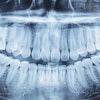
Research presented at the American Academy of Oral and Maxillofacial Radiology (AAOMR) meeting last month in Savannah, GA, disputes a suggested link between radiation from dental bitewing x-rays and the development of meningioma, a benign brain tumor.
The study, conducted by researchers from the New York University (NYU) College of Dentistry, Stony Brook University School of Dental Medicine, and Memorial Sloan-Kettering Cancer Center, is the first to evaluate bitewing exposure on both male and female CIRS phantoms and provide in-depth organ dose data, according to the AAOMR presentation.
University College of Dentistry
An epidemiological study published earlier this year in the journal Cancer prompted a flurry of media coverage and public concern because it claimed to have found a link between frequent bitewing x-ray exposure and increased risk of developing meningioma. It also prompted outcry from the oral and maxillofacial radiology community, the ADA, and the Academy of General Dentistry, all of whom questioned the study's methodology -- the findings were based on self-reporting of past dental visits from childhood through adulthood by patients who had been diagnosed with intracranial meningioma -- as well as the potential for recall bias and the fact that current x-ray exposure levels are lower than they were in the past.
"That study lacked valid statistical data on the actual patient exposure dosimetry," said Arthur Goren, DMD, a clinical associate professor at NYU in the department of cariology and comprehensive care, who presented the AAOMR paper. "You don't know what office the patient went to or what type of x-ray machines were used, so how can you pinpoint what the dose per patient was? To not have the study reviewed by a dental radiologist (prior to publication) was ludicrous."
Organ doses tested
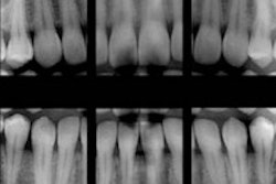
For the AAOMR study, Dr. Goren and his colleagues used a Gendex Dental Systems 765 x-ray machine to expose two anthropomorphic CIRS phantoms (30-year-old male and 30-year-old female) using the manufacturer's preset exposures for film and digital imaging and both round and rectangular collimation. All exposures were repeated 15 times, and the results were divided by 15 to evaluate the average dose per view. Thyroid collars were used during all exposures.
Optically stimulated luminescent (OSL) dosimetry was recorded at 21 head and neck structures, including the thyroid, mandible, esophagus, salivary glands, oral mucosa, and brain. The average organ dose in micrograys (µGy) was calculated based on four bitewing views.
Based on the data, the researchers found that in both the female and male phantoms, the dose to the brain was much lower than that to the other organs, Dr. Goren noted.
"Dose reconstruction techniques previously have shown that the exposure to the brain from four bitewing x-rays is approximately 0.07 mGy," he and his colleagues noted. "Here we show exposures in the 0.05- to 0.21-µGy range."
These findings call into question the Cancer study's conclusions that the development of meningiomas can be related to exposure to dental x-rays, Dr. Goren added.
"If you were going to have cancer in the brain, then you would have had it in the other organs as well," he said. "This study gives us specific data as to the dose, whereas the epidemiological study was all supposition."
Here are some other findings from the AAOMR study:
- Highest doses were seen in the glands, extrathoracic airway, and oral mucosa.
- Thyroid and other organ doses were low.
- The effective dose for the female ranged from a low of 2.6 microsieverts (µSv) (digital/rectangular) to a high of 6.0 µSv (film/round).
- The effective dose for the male ranged from a low of 3.6 µSv (digital/ rectangular) to a high of 11.5 µSv (film/round).
- Digital dose was less than film.
- Rectangular collimation dose was less than round collimation.
"We found that dentists should be using digital imaging and rectangular collimation," Dr. Goren said. "In fact, rectangular collimation should be mandatory."
A related study presented at the AAOMR meeting used juvenile phantoms and again found that digital imaging and a rectangular collimator should be used to ensure that the ALARA (as low as reasonably achievable) principle can be maximized, he added.
"Our group is the first to use juvenile CIRS phantoms with OSLs to provide organ dose data," Dr. Goren said.