The increasing adoption of cone-beam CT in dentistry has prompted much discussion of how to control the amount of radiation that patients -- particularly children in orthodontic treatment -- are exposed to.
Now researchers from the University of Würzburg have demonstrated that magnetic resonance imaging (MRI) is an effective, nonionizing option for diagnosing dental abnormalities in pediatric patients undergoing orthodontic treatment (Journal of Oral and Maxillofacial Surgery, July 2013, Vol. 71:7, pp. 1159-1169). In addition, because it is a 3D imaging modality, MRI offers more information and insight into hard- and soft-tissue dental structures than is possible with conventional radiographs.
"Radiographs are routinely used for the diagnosis of dental abnormalities and for orthodontic treatment and surgical planning," the study authors wrote. "However, radiographic images provide only limited information, especially for overlapping dental structures."
While cone-beam CT offers a 3D alternative that often becomes a necessity in complicated cases, it has limitations due to the potential radiation dose, they noted. "This limitation is especially important because most patients with dental abnormalities are children, and repetitive examinations are often required."
No sedation required
For this study, the researchers selected 16 patients with a mean age of 10.8 years from a pool of 1,500 orthodontic patients. Selection criteria were the presence of abnormalities of the size, shape, position, and number of teeth. Three of the study participants had a mesiodens, nine had supernumerary teeth other than a mesiodens, one had germination, one had dilacerations, one had transmigration, and one had transposition.
MR images were acquired using a 1.5-tesla system combined with a four-channel multifunctional radiofrequency coil array. For each scan, the patients laid on a table with the coil fixed so that its surface was slightly pressed against their lips; this provided better signal reception, according to the study authors. The children were directed to keep their mouths closed naturally and not move during each scan. No sedation was used on any patient.
Given the additional time it took to familiarize each child with the MRI system, provide ear plugs, position them on the table, and perform the scan, each procedure took about 15 minutes. Even so, "the measurement procedure and duration of 4 to 5 minutes were well tolerated by all children," the researchers wrote.
MRI yielded a clear separation between the tooth substance and the surrounding tissues, they noted. "The position and shape of malformed teeth could be assessed in all 3 spatial dimensions. The visualization software allowed viewing of the rendered structures from arbitrary angles."
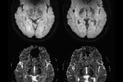
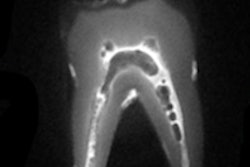
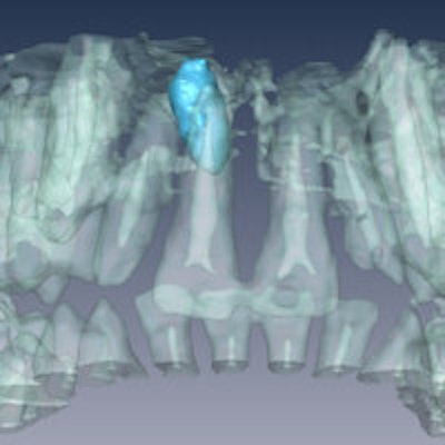
 Panoramic radiograph (above) and 3D rendering (below) of MRI data showing a mesiodens in a 12-year-old male patient. Images courtesy of Olga Tymofiyeva, PhD.
Dental abnormalities were visualized from the contrast to the adjacent signal-producing dental structure, the researchers reported. "The dental pulp, bone marrow, gingival, saliva, facial soft tissues, tongue, and palate provided a signal on clinical MRI."
The future looks bright for this imaging modality in dentistry. Compared with conventional radiographs, MRI provides the advantage of 3D morphology, which can help optimize surgical treatment and increase patient safety, they noted.
"In recent years, 3D imaging techniques have continued to evolve in dentistry beyond maxillofacial surgical planning," they wrote. "Dental MRI is the next such technique, adding a third dimension to the treatment planning to be introduced after techniques based on CT and digital volume tomography."