Dental schools in the U.S., U.K., and Australia are beginning to adopt cone-beam CT technology into their curricula, but few currently provide training on this equipment to predoctoral and undergraduate students, according to research conducted by Vijay Parashar, BDS, DDS, MDSc, an associate professor at the Midwestern University College of Dental Medicine in Arizona, in collaboration with colleagues at dental schools in the U.K. and Australia.
The results of the study, which evaluated the significance and inclusion of cone-beam CT in dental and postdoctoral residency education, were presented last month at the American Dental Education Association (ADEA) oral radiology program in San Diego. The study was conducted by Dr. Parashar; Eric Whaites, BDS, MSc, of the King's College London Dental Institute; and Paul Monsour, BDSc, PhD, MDSc, of the University of Queensland School of Dentistry.
It is a follow-on to research presented by Dr. Parashar in December at the American Academy of Oral and Maxillofacial Radiology meeting in San Diego. That study found that the majority of dental schools in the U.S. own cone-beam CT systems and have incorporated training in 3D image interpretation into their curricula.
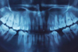
Cone-beam CT is a 3D maxillofacial imaging modality that has numerous applications in dentistry, Dr. Parashar noted during his ADEA presentation, including the following:
- Evaluating impacted teeth, supernumerary teeth, dentoalveolar fractures, orthognathic surgery, and temporomandibular joint disorders
- Diagnosing odontogenic cysts and tumors
- Endodontic evaluation of missed root canals and failed endodontic treatment
- Orthodontic treatment planning
- Airway analysis
"Traditional dental education has focused on teaching conventional 2D imaging," Dr. Parashar noted.
His study assessed the adoption of cone-beam CT technology and evaluated the incorporation of cone-beam CT teaching (taking scans and interpreting images) in dental and postdoctoral, postgraduate, and residency speciality training curricula in dental schools in the U.S. (57), U.K. (15), and Australia (7).
Dr. Parashar found that 50 dental schools in the U.S. (89%), 10 (67%) in U.K., and one (14%) in Australia presently have a cone-beam CT system. Other dental schools in Australia and the U.K . have shown interest in the technology and are in the process of actively procuring cone-beam CT equipment, he said.
However, only a small number of dental schools provide training to predoctoral and undergraduate students on how to use cone-beam CT equipment, acquire patient scan data, interpret 3D imagery, and apply implant planning software. A higher number of dental residents and postgraduates receive appropriate training in image acquisition, interpretation, and the application of implant planning software. This trend was similar across all the schools in the survey, regardless of country, according to Dr. Parashar.
"A large number of dental schools recognize the importance of teaching postdoctoral dental residents about cone-beam CT," he said. This study emphasizes the need to include cone-beam CT instruction in dental school curricula and provide dental students with more opportunities to use the technology, he added.
The audience at the ADEA meeting agreed that cone-beam CT education is highly relevant and that this study will support efforts to modify the existing curricula at their dental schools to include cone-beam CT education in the predoctoral dental curriculum.