The VELscope (LED Medical Diagnostics) is well-known as a noninvasive fluorescence device used by dental professionals to identify tissue abnormalities -- including cancerous and precancerous lesions -- in the oral cavity.
Now a new study has found that the VELscope may also be a useful aid in the surgical treatment of osteonecrosis of the jaw induced by bisphosphonates (BRONJ) (Journal of Cranio-Maxillofacial Surgery, September 4, 2013).
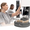
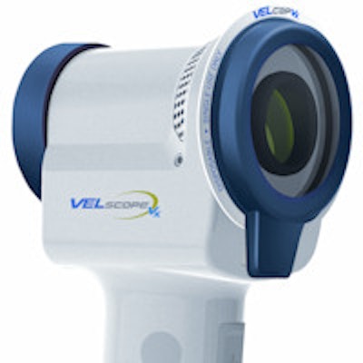
 The VELscope Vx by LED Medical Diagnostics.
"The VELscope has been widely used in the diagnosis of precancerous lesions, such as leukoplakia, carcinoma in situ, or squamous cell carcinoma of the oral cavity using autofluorescence imaging," wrote the researchers, from the University of Hamburg. "To our knowledge, there are only a few studies demonstrating the efficacy of fluorescence imaging of healthy versus necrotic bone in patients with BRONJ."
Because there are no universally accepted guidelines for surgical procedures dealing with BRONJ patients, high rates of recurrences after surgical resection have been reported, the study authors noted. They postulate that the VELscope could serve as an intraoperative guide to differentiate necrotic lesions induced by bisphosphonates from healthy bones.
They followed 20 patients for 18 months, all of whom presented to the University Medical Center Hamburg Eppendorf for initial evaluation of BRONJ. All patients had received oral or intravenous bisphosphonates for a period of nine to 84 months. Of these patients, 37 were found to have BRONJ, and 20 of the 37 were included in the current study.
Patients were included in the study if they had stage II or III BRONJ, although two patients with stage I cases also were included due to widely exposed and necrotic bone. All included patients were operated on under a special protocol; after surgical exposure of the necrotic bone, photographs were taken under normal conditions of illumination and then with the VELscope under dimmed room lights to measure the loss of fluorescence as a way to detect the presence of necrotic bone.
All patients were given doxycycline as a fluorescence marker for osseous structures. Visual fluorescence retention and visual fluorescence loss were documented, and visual fluorescence loss was considered as a positive sign for necrotic bone lesion. Osseous biopsies were taken to confirm definite histopathological diagnosis of BRONJ in each case.
The VELscope confirmed the BRONJ diagnosis in all 20 patients, the researchers reported. In 19 of the 20 patients, the VELscope was able to differentiate between healthy and necrotic bone by visual fluorescence retention and visual fluorescence loss.
"Combined with the ability of the VELscope technique to detect necrotic bone areas in patients with BRONJ by fluorescence -- as verified with the results of our study -- we share the opinion of [other studies] that fluorescence-guided bone resection offers an important and pioneering opportunity to standardize a surgical protocol in patients with BRONJ at stages II and III and in selected cases at stage I."