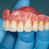
Leaded glasses and thyroid collars can effectively reduce patient exposure to radiation during cone-beam CT (CBCT) imaging without disturbing diagnostic image quality at several anatomic areas, including the alveolar processes, according to research presented at the American Academy of Oral and Maxillofacial Radiology (AAOMR) annual meeting in Chicago last month.
In a previous study, researchers from New York University, Stony Brook University, Columbia University, and Memorial Sloan-Kettering Cancer Center reported that the use of leaded glasses reduced radiation dose to the eye from a commercially available cone-beam CT system by 62% for a full field-of-view (FOV) scan and 38% for a collimated scan (Oral Surgery, Oral Medicine, Oral Pathology, Oral Radiology, and Endodontology, October 2011, Vol. 112:4, pp. 502-507).
"Because leaded glasses are relatively inexpensive and can be purchased from many different manufacturers, they should be worn by patients during all cone-beam CT scanning procedures (both limited-volume scans that do not include the eyes and full-head scans) to limit the radiation absorbed dose to the lens of the eye and reduce the risk of radiation cataract development," the study authors concluded.
In a follow-up study presented at the AAOMR meeting, the same research team studied the effect of shielding on the equivalent doses to the lens of the eye, thyroid, and other head and neck organs from a cone-beam CT system (Platinum i-CAT, Imaging Sciences International) using an anthropomorphic female phantom. All exposures were performed on one machine using five different collimation settings and preset exposure parameters. Scans were performed with and without leaded glasses, with and without a lead thyroid shield, and with and without a combination of leaded glasses and lead thyroid shield.
Each scan was repeated three times, and all radiation measurements were recorded at eight key head and neck organs using optically stimulated luminescence (OSL) dosimetry.
The researchers found that the absorbed dose to the left eye was 0.450 centi-Gray units (cGy) without leaded glasses or thyroid shield and 0.116 cGy with glasses or thyroid shield. Measurements at the right eye yielded similar values. The equivalent dose to the thyroid decreased from 63.2 micro-Sievert units (µSv) to 36.0 µSv when both glasses and shield were used. The brain, cranium, cervical spine, mandible, and parotid showed similar dose reductions, the researchers noted. Additional substantive organ dose savings resulted from machine collimation for organs located outside the direct scanning field.
Both the leaded glasses and the thyroid shield successfully reduced patient dose from cone-beam CT, the researchers noted. One problem was that the size of the leaded glasses interfered with the evaluation of certain anatomic features located between the maxillary sinus floor and the superior orbital rim, they concluded, adding that smaller glasses may ameliorate this issue.
Evaluating image quality
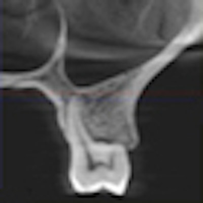
In a related study presented as a poster at the AAOMR meeting, the researchers focused on whether such shielding devices impact cone-beam CT image quality. Using the same anthropomorphic female phantom, they obtained cone-beam CT scans of radiopaque linear test objects embedded in a horizontal star pattern in the anterior mandibular floor region and a vertical parallel pattern in the left posterior alveolar processes. They also obtained full FOV cone-beam CT scans of a dry skull to evaluate the image quality at major anatomic landmarks.
','dvPres', 'clsTopBtn', 'true' );">
All scans were captured on the i-CAT Platinum system using the manufacturer's predefined settings for full field-of-view. Separate scans were acquired with and without leaded eyeglasses, and a thyroid shield was placed immediately and strictly below the mandibular base. Two oral and maxillofacial radiologists, one orthodontist, one orthodontic resident, and one radiologic technologist evaluated differences in image sharpness, image distortion, and intensity of artifacts on pairs of axial and alveolar cross-sectional images derived from volumes acquired with and without shielding devices.
The reviewers noted no perceived changes in image sharpness, distortion, or intensity of artifacts at axial levels above the thyroid shield and below the eyeglasses, according to the researchers. However, using leaded glasses and thyroid shields induced horizontal metallic streak artifacts, which resulted in dramatically decreased image quality at axial levels between the maxillary sinus floor and the orbit.
"However, [these artifacts] had no significant impact on the observers' ability to identify and evaluate major anatomic landmarks at the alveolar processes. At axial levels of the shielding devices, anatomic landmarks, such as the lacrimal duct, were obscured by streak artifacts when leaded eyeglasses were used," the researchers noted.
No significant loss of diagnostic image quality was noted at the maxillary and mandibular alveolar processes, mandibular body, and floor of the maxillary sinuses, they added.
Some surprising findings
These findings further support the use of patient shielding devices during cone-beam CT imaging, in particular when the region of interest is at the level of the alveolar processes of the jaws, according to co-author Dan Colosi, DDS, PhD, director of the division of diagnostic imaging at Stony Brook University School of Dental Medicine.
"One of the findings of our research was that even for scans limited to the maxilla or mandible, in which the eyes are not in the direct scanning field, there is some benefit to using shielding glasses in terms of further reducing radiation exposure to the eye lens," he said in an interview with DrBicuspid.com. "And when we looked at the image quality of the maxillary and mandibular alveolar processes, the use of thyroid and eye shielding did not have a perceived effect on the image quality, which we found encouraging for such clinical applications of CBCT as dental implants treatment planning."
They also found that the effects of the two shielding devices seemed to be additive in some cases, even for organs not directly close to the shielding devices, Dr. Colosi noted.
"For example, the dose to the thyroid was greatly diminished by the thyroid shield, but when we used both devices we saw an additional slight decrease in radiation dose," he said. "We haven't examined the statistical significance of this yet, but this is one area I believe would be interesting for future study."
While many states already mandate or recommend thyroid shields for many radiographic procedures, including those used in dentistry, leaded glasses are used primarily in fluoroscopy and interventional radiology, Dr. Colosi noted.
"It is certainly desirable to decrease radiation exposure to all head and neck organs, but especially to the thyroid and eye lens, which are particularly radiosensitive, without decreasing the diagnostic quality of the CBCT datasets," he added.