Ceramic mandibular veneers are likely to be as successful as veneer restorations placed on maxillary anterior teeth, according to a new study in the Journal of Esthetic and Restorative Dentistry (September 26, 2012).
"Veneers are becoming more and more important and accepted as a routine procedure in private practice," said lead study author Sven Rinke, DMD, in a DrBicuspid.com interview. "Nevertheless, data on extended veneer restoration and for veneers on mandibular teeth are still sparse."
Limited data are available from clinical studies with respect to the success of veneer placements for the restoration of mandibular incisors, and the majority of previous trials included only very few, if any, mandibular veneer restorations, the authors wrote.
Based on the current clinical data, no statement can be made regarding the clinical performance of mandibular compared with maxillary veneers. The researchers therefore conducted a retrospective evaluation of anterior ceramic veneers in the upper and lower mandible 36 months after placement.
The study included 37 patients (21 female, 16 male) who received extensive anterior veneer restorations in the maxillary and mandibular regions (teeth #6-11 and #22-27) between July 1, 2002, and June 30, 2008. The mean age of the patients at the time of insertion was 46.
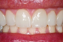
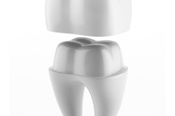
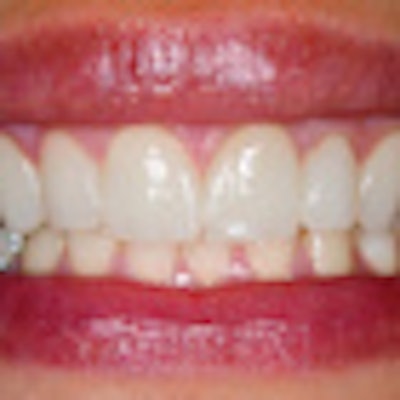
One dentist restored a total of 130 teeth where 76 veneers were placed in the maxilla, and the remaining restorations were performed on the anterior mandibular teeth. The teeth were restored with adhesively luted extensive ceramic veneers made from a heat-pressed ceramic. Adhesive cementation was performed with an etch-and-rinse adhesive and a dual-curing composite cement.
Of the 130 preparations, 73 exhibited a dentin or filling surface exposure of less than 50% of the complete preparation area, while the remaining 57 restorations were performed on preparation areas consisting of more than 50% exposed dentin or filling material.
All of the veneers were placed at Dr. Rinke's general private practice. The exclusion criteria included nonvital teeth, those with pre-existing restorations that were too large to be completely covered by the new restorations, and if large wedge-shaped defects or signs of bruxism were observed.
Teeth with one of the following indications were veneered:
- Misalignment
- Diastema
- Discolorations
- Coronal fractures
- Morphologic modifications, including prolongation of the incisal edge
- Malformations
Follow-up examinations were performed between 2009 and 2010 by a dentist different from the clinician who placed the restorations. All of the available restorations were clinically assessed using mirrors, probes, and intraoral photographs. Pulp vitality was verified with a CO2 test. Each restoration was examined for cracks, fractures, debonding, caries, and marginal discoloration.
Here are some of the key results:
- After 36 months, the survival rate was 95%.
- Reasons for failure were four ceramic fractures and one biological failure in five restored teeth.
- Of the restorations, 92% were in service without any clinical intervention and rated successful after 36 months.
- Interventions were necessary in five cases (three recementations, two endodontic treatments).
- Clinical performance was not influenced by the veneer position.
- Veneers with more than 50% of exposed dentin demonstrated a significantly increased risk for a clinical intervention, whereas no effect on the survival rate could be detected.
"After 36 months of clinical service, extensive veneer restorations made of a pressable ceramic showed a comparable survival and success rate in the upper and lower jaw," the authors concluded. "Large areas of exposed dentin were associated with lower success rates."
Although these findings were not particularly surprising, this information was missing from scientific literature, Dr. Rinke said. The information that veneers on lower incisors show the same clinical performance as veneers placed on maxillary teeth is new and can be of benefit to clinicians.
"These findings give evidence for a broadening of the field of indications for veneers," Dr. Rinke added.
However, the study authors noted that their study is limited because of the relatively short mean observational period of 36 months.
"Because the number of participants who were available for follow-up examinations decreased over time and all of the restorations were performed by a single operator, these findings should be interpreted with caution," the authors concluded.