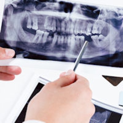
When examining periodontal pocket depth, soft-tissue measurements can be affected by the examiner's skill and technique. However, an indirect radiographic method may be the best way to determine periodontal bone levels, according to a new study in BMC Oral Health.
Registrations made on radiographs are influenced by the techniques by which the images, as well as the measurements, are obtained (registration is defined as the precise alignment of two images of the same object). A long cone "paralleling technique" with a standardized receptor holder is recommended for obtaining radiographs, and it has been suggested that the direct method (measured in millimeters) should be used in clinical trials when investigating the susceptibility of different teeth to periodontal breakdown.
 Hans Preus, DDS, PhD, professor, Department of Periodontology, University of Oslo, Norway.
However, researchers have warned that image distortions of the object (tooth) appear if there is deviation from the paralleling technique.
Using a direct method of measurement was markedly biased when the angle between the x-ray beam and the sensor was 30°, while the indirect technique was not biased, Norwegian researchers found.
"[The indirect method] is an easy way to eliminate/reduce the errors that comes with different angulation of the x-ray cone between two radiographs obtained at two different time points," lead study author Hans Preus, DDS, PhD, told DrBicuspid.com. Dr. Preus is a professor in the department of periodontology at the University of Oslo in Norway.
Evaluating periodontal bone levels
Periodontal destruction is usually described in periodontal pocket depth (PD) in millimeters and clinical attachment level. A decrease or increase in the alveolar bone level at a given site over a period of time may be regarded as progression or regression of the disease process at that site.
However, these soft-tissue measurements are considered inaccurate parameters, according to the study authors, and reflect an unpredictable combination of gingivitis and periodontitis, while also being affected by an examiner's skill, reliability, and technique. For these reasons, radiographic examination is considered a more reliable method of measurement.
In longitudinal clinical studies, the reproducibility of registrations is critical, and the distortion of imaged teeth and surrounding bone on radiographs will produce unreliable measurements of bone levels and tooth length. Consequently, indirect imaging creates incorrect impressions of bone loss or gain over time. Researchers need new techniques that could correct such distortions, the study authors noted.
This study compared two evaluation methods: an indirect radiological examination technique for measuring radiographic bone level and a technique that used direct millimeter measurements. The researchers wanted to see if examiner bias could be substantially reduced (BMC Oral Health, September 8, 2015).
Experimenting with mandibles
In this study, researchers used seven human dry mandibles with 20 loose teeth to control the angle between the receptor and x-ray beam, which was perpendicular to the central beam. None of the loose teeth or surrounding bone in the mandibles showed signs of severe periodontal disease since the mean distance from alveolar crest to the cementoenamel junction was 1.7 mm.
"Severe" or "extensive" periodontitis is reflected by clinical attachment loss of more than 5 mm to 7 mm, which corresponds to a radiographic bone level of greater than 7 mm.
Since the dry mandibles did not show such degrees of bone loss, an artificially reduced bone level was created by placing hard wax in the 20 sockets and replacing the teeth into the wax, allowing a stable marginal bone level of approximately 7 mm.
In one set of images, the mesial and distal radiographic bone levels were obtained by direct measurements using traditional software. In the other, an experimental software plugin was applied, which produced a length-adjusted radiographic bone level. A total of 40 sites on 20 radiographs obtained at each angle (0° and 30°) were read twice with both the direct and indirect techniques.
Significant differences in measurements
When the mean radiographic bone level measured at 0° was 7 mm, the corresponding mean bone level measured at 30° was 7.8 mm. The 11.4 % increase was statistically significant (p = 0.032). The study showed that tooth length was significantly increased from one image to the next as the angle increased from 0° to 30° (p < 0.001).
The reliability of tooth length measured by the direct method was 0.96 (95% confidence interval [CI]: 0.94 to 0.98), compared with 0.98 (95% CI: 0.96 to 0.99) using the indirect method, the study showed. The reliability of radiographic bone level when measured by the direct method was 0.86 (95% CI: 0.81 to 0.89) and 0.94 (95% CI: 0.90 to 0.97) using the indirect technique.
"The results from this study was absolutely expected, since it is a natural consequence of angulation-induced errors," Dr. Preus said.
If the objects being measured are larger than 2 mm, the indirect technique is a useful and reliable tool, the researchers found. It is also faster to use and easier to assess, they noted.
Applications for indirect technique
In addition to periodontal measurements, the indirect technique could also be used in oral surgery and caries and endodontic evaluations, according to the study authors.
While the findings are particularly relevant to ongoing longitudinal studies, the authors noted that with the ongoing development of digital radiographic techniques, it may eventually be useful to clinicians as well.
"It is a digitalized and versatile version of the Schei ruler and Björns technique, and is at present more important to research than clinics," Dr. Preus said. "But in research, it is a very useful tool, since radiographic images of periodontal destruction have been used less than soft-tissue measurements like pocket depth and clinical attachment level."