X-rays may weaken bond strength of adhesives to dentin, researchers from the State University of Campinas report in the current Journal of Adhesive Dentistry (November 2009, Vol. 11:5, pp. 355-360).
They used flat dentin surfaces on human molars and built them up with Z250 composite (3M ESPE), into cylinders using three different adhesive systems: a two-step etch-and-rinse (Single Bond 2 – SB2, 3M ESPE), a two-step self-etching (Clearfil SE Bond – CSE, Kuraray), or a singlestep self-etching (Adper Prompt – ADP, 3M ESPE).
They assigned the specimens to four groups of 10 specimens each, according to the X-ray dose: 0 (the control), 5 Gy, 35 Gy, or 70 Gy.
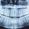
Four full-mouth bitewing radiographs with D speed film and round collimation exposes the patient to about 17 micro Gy (17/1,000,000 of a Gy), according to Principles of Dental Imaging (Baltimore: Williams & Wilkins, 1997).
The researchers directed radiation onto the surface of the resin cylinders.
After 24 hours, they tested microshear and classified the failure modes under 200X magnification.
They found that in general, the greater the radiation dose, the weaker the bond (R2 ≥ 0.905). The type of adhesive also affected the strength. For SB2, the control's strength was greater than the specimen than underwent 5 Gy, which was equal to the specimen that underwent 35 Gy, which was greater than the specimen that underwent 70 Gy.
Similarly, for CSE the strength of the control = 5 > 35 = 70. And for ADP, the control = 5 = 35 = 70. Generally, SB2 > CSE > ADP.