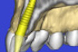
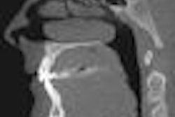
Cone-beam CT (CBCT) enables better visualization of miniscrew implants when they are used as temporary anchorage devices (TADs) for orthodontic treatment, according to a study in the American Journal of Orthodontic and Dentofacial Orthopedics (February 2010, Vol. 137:2, pp. 166-167).
The study, conducted at the University of Texas Health Science Center in Houston, evaluated the location of TADs placed during orthodontic treatment, with particular emphasis on implant placement with regard to the surrounding dentoalveolar structures.
Cone-beam CT scans were taken before and after placement of the TADs over a six-month period as part of a routine clinical protocol. Thirty-five TADs (19 in the maxilla, 16 in the mandible) were evaluated with regard to placement site, length of the TAD in the alveolar bone, amount of contact with the periodontal ligament, and inter-root distance between TADs.
The researchers found that mean lengths of the TADs in alveolar bone were 5.29 ± 1.39 mm in the maxilla and 4.60 ± 0.86 mm in the mandible. The amounts of contact with the periodontal ligaments were 2.54 ± 0.81 mm (n = 13) in the maxilla and 2.72 ± 0.49 mm (n = 10) in the mandible. The inter-root distance measurements were 2.78 ± 0.76 mm (n = 15) and 5.19 ± 4.42 mm (n = 16) in the maxilla and the mandible, respectively.
"Three-dimensional cone-beam computed tomography technology allows better visualization of TAD placement," the authors wrote. "Clinicians can expect 71.2% of the length of the screw section of the TAD to be embedded in the alveolar bone; the percentage is often higher in the maxilla than in the mandible."
Of the 35 TADs, 65.2% were in contact with the periodontal ligament, they added, leading them to conclude that there appears to be more space for TAD placement in the mandible than in the maxilla.
Copyright © 2010 DrBicuspid.com