In response to the increasing utilization of cone-beam CT for a broad range of diagnostic indications and growing concerns about radiation dose exposure, digital imaging vendors are working to develop new products that address evolving dental needs.
"Demands for imaging in dentistry have changed," said Jeffrey Brooks, DMD, an assistant professor in the department of oral and maxillofacial surgery at the University of Tennessee College of Dentistry and clinical director of 3D imaging for Carestream Dental. "Cone-beam CT was originally used for implant planning and pathology. But in the last five to seven years that has completely changed, and its uses have grown dramatically in dentistry."
— Jeffrey Brooks, DMD, Carestream
Dental
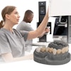
This evolution prompted Carestream to develop the CS 9300-1 extraoral imaging system. Introduced this month at the California Dental Association spring meeting in Anaheim, the CS 9300-1 features up to seven selectable fields-of-view (FOVs), ranging from 5 x 5 cm to 17 x 13.5 cm, enabling practitioners to focus on a specific region of interest by collimating the field-of-view.
Not only does this broaden a practitioner's ability to use a single system for multiple applications, it limits patients' radiation exposure, Dr. Brooks noted.
"Dental practitioners no longer have an either/or situation when it comes to 3D imaging -- that is, whether to take a full field-of-view image or not," said Robert Patrick, director of U.S. product line management at Carestream Dental.
Multispecialty applications
The CS 9300-1 allows the practitioner to focus on the specific FOV that is optimum for a particular diagnosis, explained Dr. Brooks, who has worked with the system for several months at the University of Tennessee.
"In endodontics, for example, when the focus is on a small area such as a single tooth, this system allows the practitioner to collimate the FOV down to a 5 x 5-cm image and still retain a resolution of 90 microns," he said.
The CS 9300-1 also offers a large FOV for orthodontic and oral and maxillofacial surgical applications, such as 10 x 10-cm and 8 x 8-cm FOVs that allow for dual-arch implant planning and 10 x 5 cm for single-arch implant planning, Dr. Brooks added. There is also a FOV that accommodates the evaluation of temporomandibular joints bilaterally.
 |
| Click here to enlarge this image. |
| The CS 9300-1 features up to seven selectable fields-of-view for 3D images, ranging from 5 x 5 cm to 17 x 13.5 cm. Image courtesy of Caresteam Dental. |
"So you can see that there is a tremendous amount of flexibility in selecting the FOV for any specific diagnosis, which is very important for reducing radiation dose," he said. "Clinicians do not like having to scan more area on a patient than is diagnostically needed."
The CS 9300-1 also offers 2D digital panoramic imaging with variable focal trough technology. In addition, its fast scan times help lower the risk of retakes due to patient movement, according to Dr. Brooks.
"It is important not only to collimate down to any specific area but also to have a 2D modality because panoramic imaging is the workhorse in dentistry," he said. "So the ability to have the same unit have multiple functionalities is very important. So what sets this unit's 2D panoramic capabilities apart is also the variable focal trough functionality."
The CS 9300-1 also comes preinstalled with Carestream Dental's CS 3D imaging software, Dr. Brooks noted.
"The big bonus here is the fact that you have an all-inclusive imaging system that gives practitioners the ultimate flexibility in terms of 3D and 2D imaging," he said. "That is where we've been somewhat restricted with other units because we have been somewhat forced to use the FOVs that were presented in those units, as opposed to having the flexibility in a single unit to position a patient, focus in on the area only that needed to be acquired, and then view and manipulate it using the software."
Allan Farman, BDS, MBA, PhD, DSc, a professor of radiology and imaging science at the University of Louisville in Kentucky and president of the American Academy of Oral and Maxillofacial Radiology, hasn't worked with the CS 9300-1 but said the development of this type of system is a logical next step.
"As cone-beam CT enters the mature development phase, it should be expected that systems will provide more sophisticated physical collimation mechanisms to restrict dose," he told DrBicuspid.com. "Exposure to tissues outside the region of interest should be protected to the extent possible from radiation. The CS 9300 fits with this better class of cone-beam CT instruments with respect to opportunities to collimate; however, my definitive opinion will need to wait for confirmation of the actual levels of dose for specific tasks versus the diagnostic image quality as demonstrated by research conducted independently of Carestream."
While the CS 9300-1 could be a step forward for Carestream's product line, "this product is not unique in its ability to be collimated," Dr. Farman added. "Perhaps all maxillofacial cone-beam CT units should be held to this standard before approval for use in the future."