Dental practitioners and oral and maxillofacial radiologists need to be aware of the extent of incidental findings in the maxillofacial region that can be detected by cone-beam CT (CBCT), according to a study in Diagnostic Interventional Radiology (September 29, 2011).
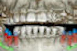
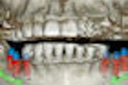
Researchers from Ataturk University in Turkey examined cone-beam CT images of 207 patients (129 females and 78 males). The sample consisted of 85 temporomandibular joint (TMJ) disorder patients, 45 paranasal sinusitis patients, 30 obstructive sleep apnea syndrome patients, 15 implant patients, and 32 others.
The overall rate of incidental findings was 92.8%. The highest rate of incidental findings was in the airway area (51.8%), followed by impacted teeth (21.7%), TMJ findings (11.1%), endodontic lesions (4.3%), condensing osteitis (1%), and others (2.9%). The airway incidental findings included mucosal thickening (21.3%), deviation of the nasal septum (12.6%), conchal hypertrophy (11.1%), bullous concha (3.9%), and retention cysts (2.9%). The impacted teeth consisted of third molars (18.8%) and canines (2.9%). The incidental findings for the TMJ patients were erosion of the condyle (4.8%), osteophytes (3.4%), and bifid condyle (2.9%).
"Oral radiologists should be aware of possible incidental findings and should be vigilant about comprehensively evaluating possible underlying diseases," the study authors concluded.


