Dental researchers are making progress in their quest to develop a practical magnetic resonance imaging (MRI) device that can be used in the operatory for hard- and soft-tissue analysis and diagnosis.
In a technical paper published in Magnetic Resonance in Medicine (July 30, 2013), scientists from the University of Minnesota Center for Magnetic Resonance Research outline their efforts to optimize the MR signal from the teeth and supporting structures -- an important step in improving overall image resolution, particularly with hard tissue.
"Dental MRI has the potential to be even more informative than x-ray imaging techniques by visualizing, noninvasively and simultaneously, both hard and soft tissues in all three spatial dimensions," they wrote. "However, clinical MRI has yet to attain the resolution of cone-beam computerized tomography imaging."
At present, at least four MRI methods can be used to obtain images of densely calcified dental tissues, the researchers noted: ultrashort echo times (TE), sweep imaging with Fourier transformation (SWIFT), zero TE, and combined pointwise encoding time reduction with radial acquisition (PETRA).
"These methods now make it feasible to image these tissues, but further RF [radio frequency] coil refinement is still needed to optimize the MR signal from the teeth and supporting structures," they wrote.
The core of the issue involves overcoming patient motion, including swallowing. This requirement that can only be met "when the coil positioning does not create excessive discomfort for the patient," they explained.
','dvPres', 'clsTopBtn', 'true' );" >
Noting that the most comfortable coil position -- the occlusal plane -- has not been seriously considered in previous dental MRI studies, the researchers set out to demonstrate the advantages of using a loop coil in the occlusal position for dental imaging.
"In the occlusal position ... the sensitive volume of the coil encompasses the most important dental structures -- the teeth and their supporting structures -- while uninteresting tissues containing much higher proton density (cheeks, lips, and tongue) are outside the sensitive volume," they wrote.
For their experiments, they constructed a single loop using 10-mm-wide copper foil; the shape and size were chosen to fit the average adult maxillary arch (about a 25-mm radius). They covered the foil with sticky foam for comfort.
','dvPres', 'clsTopBtn', 'true' );" >
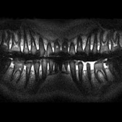
All phantom and in vivo images were acquired using a 4-tesla MRI system equipped with a Varian DirectDrive console. The in vivo images were obtained from a normal adult volunteer. The coil was inserted in the occlusal position and isolated from intraoral structures and saliva. The patient was placed in a supine position with his head in a holder specifically designed to restrict head motion. The researchers chose the SWIFT method for all imaging.
The resulting images are the first MRI panoramic images to demonstrate high nominal resolution (0.3 mm3), the researchers noted. The images show that the teeth and jaw bones are well positioned within the sensitive volume of the coil, while the signal from the cheek and tongue yield low intensity, they added.
"The presented images and simulated data show the advantages of using a coil in the orthogonal orientation for dental applications," they wrote. "The transverse components of the B1 field of a surface coil can effectively be used for imaging of teeth and associated structures."