Finding vertical root fractures can be tricky. As digital imaging technology improves, clinicians and researchers are fine-tuning the best approach for radiographically detecting vertical root fractures, and there is plenty of room for improvement at present.
Currently, it requires a combination of clinical signs and periapical radiography as the first imaging modality employed, noted Brazilian researchers from the division of oral radiology at the State University of Campinas Piracicaba Dental School and the department of clinical dentistry at the Federal University of Espírito Santo Faculty of Dentistry. But there are significant limitations to an approach that creates two-dimensional representations of dentition.
Multiple radiographs may need to be taken to find these fractures, and unless the beam passes along the line of the fracture, it may not be revealed at all, they noted in their study published in the International Endodontic Journal (July 7, 2014).
Previous research has examined the use of software enhancement filters when diagnosing caries with digital images, but vertical root fractures have received little attention, and the studies the researchers found employed charge-coupled device (CCD) digital systems instead of phosphor storage plates (PSPs). In the present study, the researchers compared the diagnostic accuracy of different digital enhancement filters available on the Digora Optime digital system (Soredex) using PSPs while attempting to detect vertical root fractures.
They were straightforward in their conclusion: "Digora Optime images with the Sharpen filter had the best performance-related indices for visualizing root fractures in vitro," they wrote. "The Sharpen filter had the highest sensitivity and accuracy."
Interestingly, the researchers found "little information" from the manufacturer about each of the enhancement filters: Negative, 3D Emboss, Shadow, and Sharpen. Negative simply swaps blacks in the image to white and vice versa.
"Both 3D Emboss and Shadow can be used to visualize paths of structures that pass through dark and light regions of an image, such as endodontic files for length measurement, or root fractures and similar pathology," the researchers explained. "A residual portion of grayscale remains when using the Shadow filter."
The Sharpen feature is designed to enhance subtle aspects of an image. It uses an algorithm and computer-assisted manipulation of the image to group high-contrast pixels together in a submatrix, bringing forward areas of the image produced that may not be as noticeable otherwise.
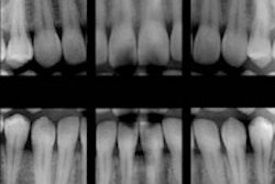
To test these filters, the researchers gathered 40 extracted single-rooted human teeth for their study, none of which had root fractures, root resorption, endodontic treatment, obliterated root canals, pulp calcifications, or supernumary roots or canals. Twenty were given vertical root fractures that had no division in two fragments with an Instron machine. The fracture was confirmed with a transillumination view. All roots were placed in the premolar region of a dry human mandible socket.
The imaging phase of the study took place next, using the Digora Optime system with a spatial resolution of 1270 dpi. They radiographed the specimens with a GE 1000 x-ray unit (General Electric) with a 2.4-cm acrylic plate placed in front of them to simulate soft-tissue attenuation. Using one vertical angulation, 0°, and three horizontal angulations, -30°, 0°, and +30°, the researchers produced three radiographs for each specimen. This was repeated five times with an original image for each of the four filters: Sharpen, Shadow, Negative, and 3D Emboss.
After completing this process with all 40 specimens for a total of 600 images, they were randomized and blindly evaluated by three oral radiologists at a rate of 20 per day. They used a scale of 1 through 5 to score each image with 1 representing the absence of a root fracture, 3 representing uncertainty, and 5 indicating the presence of one. They were not allowed to adjust the brightness, contrast, or zoom to isolate the influence of each filter.
The researchers explained that they broke down their data into three areas:
- Sensitivity, for correctly identifying vertical root fractures
- Specificity, for correctly identifying the lack of vertical root fractures
- Accuracy, or the proportion of correctness
The Sharpen filter yielded the highest sensitivity and accuracy, while the Negative one had the highest specificity. The Negative filter also had the lowest sensitivity, however, while Shadow had the lowest specificity and 3D Emboss had the lowest accuracy.
The researchers calculated the receiver operating characteristic (ROC) curve to gauge the sensitivity-specificity relationship in the diagnosis of each group. The Sharpen filter proved to be the best when the areas under the ROC curves (Az) for each of the five settings were compared and analyzed. It produced significantly higher Az values (p < 0.05) than the other filtered and original images.
"Unlike previous studies, a significant improvement in the detection of vertical root fractures was observed using the Sharpen filter," the researchers wrote.