As the success of dental implants and implant restorations depends on correct diagnosis and treatment planning, a reliable, cost-effective method of creating a reliable 3D surgical guide is crucial.
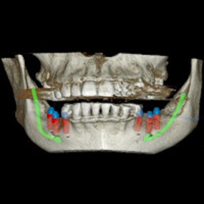
Now a new study reports that stitched small field-of-view (FOV) cone-beam CT (CBCT) images appear accurate and reliable enough for the fabrication of these surgical guides. These stitched images can be used to measure important features of the mandible and create a reliable 3D surgical guide.
The study, which was published in Imaging Science in Dentistry, found that the mean difference between stitched small FOV CBCT measurements and the reference standard of anatomic linear measurements was 0.34 mm. As most clinical dental procedures measure margin of error by 1 mm, this difference is not a clinically relevant one, according to the authors.
"Stitched composite 3D imaging appeared accurate and reliable for diagnostic purposes," wrote lead author Nicholas Egbert, DDS, and colleagues. "The miniscule differences between the control measures and those of the stitched data sets allow image-guided implant surgical stents to be fabricated from such datasets" (Imaging Sci Dent, March 2015, Vol. 45:1, pp. 41-47).
3D stitching vs. digital caliper
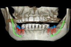
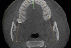
The researchers set out to evaluate whether stitched small FOV images could be reliably and accurately used for surgical dental implant guides. 3D stitching is a new software mode that combines two or three small FOV images to create a composite 3D image.
3D stitching has several advantages for dentists and endodontists, as the software is flexible and has clearer image quality than large FOV images, according to the researchers.
But none of those advantages are important if the stitched images aren't accurate and reliable enough for use as a clinical guide. So Dr. Egbert and colleagues put three reference points and 10 additional markers on a cadaveric mandible, and they measured the difference in points with a precision sliding digital caliper, which served as a control.
They then scanned the mandible and composited the images using the latest version of Kodak 9000 software. The researchers took the same measurements with the small FOV CBCT stitched images, and compared them to those acquired with the digital calipers.
The researchers found that the mean difference between the calipers and the stitched small FOV CBCT measurements was 0.34 mm, and the mean standard deviation was 0.30 mm. They determined this difference to be clinically insignificant because the human eye and hand cannot detect submillimeter differences during surgeries.
"There was no systematic bias between the differences in the observations," the authors wrote. "Each measurement appeared to be as good as the other. The differences between the control measurements and the CBCT measurements were acceptable within the parameters of this study."
How to ensure reliable measurements from stitched images
However, the authors did note that CBCT operators should pay attention to any significant discrepancies in measurements between anatomical markers and their reconstructed images.
"If significant discrepancies in measurement occur, the scan or stitched volumes might be inaccurate, causing the subsequent guide to be inaccurate," Dr. Egbert and colleagues wrote. "In this situation, guided surgery should be aborted."
The authors also noted that operators can include a radiographic guide before taking the scan. The resulting measurements can then be used to determine the accuracy of the implant surgical stent.