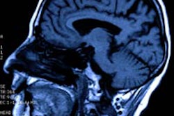
A preliminary study suggests that a magnetic resonance imaging (MRI) technique called SWIFT (sweep imaging with Fourier transform) can provide a 3Dl assessment that may aid in detecting involvement of the mandible by oral cancer, according to a report in the September issue of Archives of Otolaryngology -- Head and Neck Surgery (September 2011, Vol. 137:9, pp. 916-919).
Advanced squamous cell carcinoma that arises in the oral cavity frequently invades the mandible, according to the study authors from the University of Minnesota. Treatment may or may not necessitate removal of the mandible.
"Unfortunately, detecting bone invasion prior to surgery is often difficult using currently available imaging techniques," they wrote.
Determining mandibular invasion with a high degree of accuracy before surgery might allow the surgeon to contain the cancerous cells, prevent unnecessary mandible removal, and aid in planning for reconstruction, they added.
Although multiple imaging techniques -- primarily CT and MRI -- have been used preoperatively to assess mandibular invasion in oral carcinoma, "these techniques may not always provide a clear and accurate assessment of tumor infiltration into the mandible," the researchers wrote.
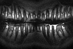
These limitations prompted them to investigate using SWIFT MRI to image mandibular invasion by squamous cell carcinoma. Participants in the descriptive case study were patients with oral carcinoma who underwent segmental mandibulectomy at a tertiary academic institution. The researchers used a 9.4-tesla MRI system (Varian Medical Systems) to examine two specimens from each patient for cortical and medullary invasion by cancer cells. Histologic sections were compared with the images obtained by the SWIFT technique.
Images produced by the SWIFT technique with in vitro specimens were of sufficient resolution (156 to 273 µm) to accurately depict tumor invasion of cortical and medullary bone, the researchers reported. Evidence of mandibular invasion with tumor was found in both specimens by histopathology. There was also a high degree of correlation between the MRIs and histopathologic findings.
"This preliminary report demonstrates that the SWIFT imaging technique has the capacity to show fine details of intramandibular anatomy," the study authors concluded. "Furthermore, the correlations between the histologic and MR images of these two specimens clearly show malignant invasion that has not been previously demonstrated with MR techniques. The data described in this report suggest that MRI has a great deal of potential in accurately determining bone invasion preoperatively."