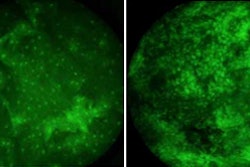
Autofluorescence imaging and spectroscopy can assist clinicians in identifying early oral cancer and advanced dysplasia in high-risk individuals, according to a study presented last week at the International Association for Dental Research meeting in Brazil.
In an ongoing clinical protocol, researchers from the University of Texas MD Anderson Cancer Center, Rutgers University, Rice University, and University of Texas Dental Branch at Houston obtained autofluorescence imaging and depth-sensitive spectroscopy data of patients with potentially malignant oral lesions and/or a history of oral cancer.
To date, more than 125 patients have been enrolled and imaged, over 60 have been imaged at least twice (median four-month intervals), and more than 80 sites have correlating pathologic data, the researchers reported. More than 10 patients have been diagnosed with cancer after enrollment.
Comparisons of clinical impression using only white light, clinical impression with white light and autofluorescence images, and quantitative autofluorescence data with clinical outcome and available histologic data suggest potential efficacy of additional autofluorescence diagnostic information, the researchers reported.
"Initial evaluation of an ongoing clinical surveillance study suggests that autofluorescence imaging and spectroscopy can provide qualitative and quantitative information to assist clinicians in the difficult clinical challenge of identifying early oral cancer and advanced dysplasia in high-risk individuals," they stated.