WASHINGTON, DC - Contrary to decades of tradition, dentists should leave carious dentin untouched in many lesions, researchers said Wednesday at the American Association for Dental Research (AADR) annual meeting.
For years, dental professors have taught their students to remove all infected tissue from cavities and carefully shape the preparation before placing a restoration, said Edwina Kidd, B.D.S., F.D.S., Ph.D., D.Sc., a former professor at King's College London.
"Does this fit with knowledge?" she asked. "I think the answer may be 'no,' which means I've been teaching unsubstantiated rubbish for 30 years."
James Summitt, D.D.S., M.S., chair of restorative dentistry at the University of Texas at San Antonio agreed, citing multiple studies that suggest sealing over a lesion can arrest its progress -- including a 10-year randomized trial in which a team led by Eva Mertz-Fairhurst, D.D.S., of the Medical College of Georgia pitted minimally invasive techniques against traditional ones and found the minimal approach superior (Journal of the American Dental Association, January 1998, Vol. 129: 1, pp. 55-66).
To explain why orthodox tenants of dentistry may be wrong, Dr. Kidd pointed to new findings about the microbiology of caries. "Understanding of the caries process is essential to understanding the question," she said. "With regular disturbance of the biofilm, the caries can be arrested. This arrest can occur at any stage. The white spot lesion can be arrested. And the large cavitated lesion can be arrested because that lesion is open and accessible to plaque removal."
On the other hand, placing a filling does no good if the patient isn't brushing properly, Dr. Kidd said, showing a slide of caries all around a restoration.
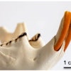
The same is true of root caries, she added, showing slides in which active caries had halted under the assault of toothbrushes and fluoride. The dentin typically becomes hard, dry, shiny, and dark as the caries stops, she said.
When to fill
So when should restorations be placed? "When the patient can't access the plaque," Dr. Kidd said.
For example, lesions that widen as they go deeper are too difficult to clean, she said, comparing them to a British marmite jar whose mouth is smaller than its body. The patient's brush just can't get past the overhanging enamel to disrupt the biofilm inside.
— Edwina Kidd, B.D.S., F.D.S., Ph.D., D.Sc.
The same is true of many interproximal lesions. "The most fastidious flosser couldn't clean plaque out of the hole," Dr. Kidd said. "They would only gloss over the surface."
In deciding whether to fill, she recommended using radiography to see how far a lesion has progressed into the dentin. Clinicians can use a "watchful waiting" approach for lesions that are just slightly into the dentin and might not become cavitated, she said.
For interproximal lesions, rubber orthodontic rings can separate teeth, Dr. Kidd said. About 48 hours after placement, the ring can open enough space for a clinician to gently probe for a break in the enamel.
But even in some of these cases, a clinician might get away with sealing over the lesion, she said. For deciduous teeth, overhanging enamel can be removed to allow cleansing -- rather than filling -- of the lesion. "Which approach sends the message that the patient is in charge?" she asked.
But sometimes even that much drilling is too much, Dr. Kidd said. "Could one even do nothing, simply relying on the deciduous nature of the teeth?" She cited a couple of studies finding that caries progresses so slowly that more than 80% of carious deciduous teeth fell out before causing any trouble (British Dental Journal, July 2002, Vol. 193:2, pp. 99-103, 219-223).
Some of the typical indications for replacing restorations on recurrent caries may be wrong as well, according to Dr. Kidd. "Recurrent caries is primary caries at the margin of a filling -- it's as simple as that," she said, so it should be treated the same way -- cleaned rather than filled, when possible. Most important: Staining around the margin doesn't indicate new infection.
More accurate indications of when to replace a restoration are x-rays showing lesions that can't be cleaned, or a ditch in the margin large enough to be probed with an explorer, she said.
Buried alive
And even when a restoration is needed, it may be sufficient to seal over the cavity without removing the infected tissue, said Dr. Kidd, citing four randomized, controlled trials of this approach. In two of them, the investigators re-entered the lesions to see what had happened under the filling, and the results looked promising, she said.
She also cited a study of the Hall technique, in which a preformed metal crown is placed over an unprepared molar. The procedure is less traumatic for the patient -- typically a child. A randomized trial led by Nicola Innes, B.M.Sc., B.D.S., a general dentist near Dundee, Scotland, in BMC Oral Health (December 2007, Vol. 20:7, p. 18) found that this type of restoration had no more failure than traditional restorations for the same type of lesion. (The high occlusion self-corrects, Dr. Kidd said.)
Under the restoration, sclerosis of the tubules and tertiary dentin reduces the permeability of the dentin, entombing the organisms under the restoration, said Dr. Kidd, citing her own study among several others, especially a study in Caries Research (October 2009, Vol. 43:5, pp. 454-358) by researchers from the University Center of Maranhão in Brazil. In the changed environment under the filling, the bacteria become less erosive, they found.
Dr. Kidd finished up with an analogy. "The apparent irrelevance of infected dentin is biologically logical if it's accepted that the caries process occurs in the biofilm and its reflection is the lesion in the dental hard tissues," she said. "Let me put it this way. As I look at my reflection in the mirror, I can muse on the ravages of time upon my face. It may be that a face lift would help. But what would not help would be to take a hammer and smash the mirror. The disappointing reflection would be in shards at my feet, but the real me would still be here."
More considerations
For his part, Dr. Summitt offered a decision tree using a more extensive set of criteria to consider in deciding whether to fill a lesion. His criteria:
- The extent of dentin involvement
- The patient's caries risk
- Keeping food out of the lesion
- Preventing tooth movement
- Reducing sensitivity
- Aesthetics
The patient's behavior has to be taken into consideration, Dr. Summitt said. Sometimes a filling is needed even in a lesion that can be cleaned because the patient just won't clean it, he said.
Not everyone in the audience was willing to swallow the idea of simply sealing over carious dentin. Edward Lynch, M.A., B.D.Sc., a Queen's University professor of restorative dentistry, cited some studies suggesting that sealing doesn't stop caries. In particular, he pointed to one led by Jan Poorterman of the Academic Centre for Dentistry Amsterdam, that found caries progression in 70% of sealed occlusal lesions (Caries Research, January-February 2003, Vol. 37:1, pp. 29-33).
"The jury is still out," Dr. Lynch said. "The evidence is mixed, and we all know that restorations can leak. So what I do is take a pharmaceutical approach, using ozone or photoactive disinfection." But he, too, touches lightly with the bur.
He promised more details in his own forthcoming AADR presentation.
Copyright © 2010 DrBicuspid.com