A method of using calcium phosphate solutions to remineralize carious lesions in dentin has had promising results in a recent study, dramatically decreasing the time needed for remineralization.
In the study performed at the University of California, San Francisco (UCSF) and presented at the International Association for Dental Research (IADR) conference in March, researchers performed a pretreatment with fluoride solutions on dehydrated and demineralized dentin and found that the process may significantly alter the remineralization process from several days down to just minutes.
The acceptance of remineralization as a treatment approach has been hampered by the length of time needed to get significant results. "We were thinking about how to accelerate the process, and that's where the fluoride treatment comes in," explained Stefan Habelitz, PhD, an associate professor in the department of preventive and restorative dental sciences at the UCSF School of Dentistry who designed the study and supervised Paul Hsaio, a dental student leading the project.
But the team also sought to functionally remineralize dentin -- "to restore the mechanical properties of the tissue by reintroducing the minerals," Habelitz said. "Because in dentin, if you don't follow certain procedures, you can incorporate minerals into the lesion but it doesn't recover the properties, it only recovers it to a small degree."
The PILP system
To achieve this, the team employed the polymer-induced liquid precursor (PILP) system created by Laurie Gower, PhD, an associate professor in the department of materials science and engineering at the University of Florida in Gainesville.
— Stefan Habelitz, PhD, University of
California, San Francisco
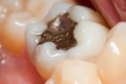
"In a carious lesion where the bacteria attack enamel and dentin, the mineral is dissolved surrounding the fibril but also inside it," Habelitz said. "When you expose the demineralized tissue to a calcium and phosphate solution, mineral formations only occur around the fibril. The solution usually does not enter the fibril."
Remineralization takes place, but the dentin does not have as much strength as it would if minerals were forming in the fibrils. However, in the PILP system, polyaspartic acid delivers calcium phosphate to the fibrils and releases it inside the collagen fibril so minerals form within them.
"Having that PILP system gave us an opportunity to fully recover the tissue, and then we looked at what it would do to a carious lesion," Habelitz said. The drawback was that it remineralizes too slowly. "Basically, it would almost take a year to remineralize a natural lesion with that method as it is right now," he said.
For the IADR study, the team used a low-speed Buehler saw and a diamond blade to cut extracted human molars. Next, the teeth were polished with a strip grinder, adhesive polishing disks, and diamond polishing slurries. The triple-polished surface provided a flat, even reference.
Next, the dentin disks were cut into 3 x 6-mm2 dentin and covered with nail varnish -- "Revlon cherry color red, I believe," Habelitz quipped -- leaving a 3 x 3-mm2 window on the occlusal surface.
"It's a very nice reference layer for comparing the mechanical properties in the natural tissue versus the demineralized and study also how quickly the mineral recovers," Habelitz said.
To make artificial caries in the samples, the researchers applied 0.5 molar (M) acetic acid with calcium and phosphate at pH 5 for 66 hours, resulting in a lesion depth of 100 µm. The lesions were completely dehydrated, then immersed into different sodium fluoride solutions for one to two minutes and dried in an incubator at 37° C for one hour.
They then immersed the specimens into pH 7 metastable remineralization of 6 mM of calcium chloride (CaCl2) and 3.8 mM of monopotassium phosphate (KH2PO4) or 8 mM CaCl2 and 5.0 mM KH2PO4 for one hour at room temperature. The control group did not receive fluoride treatment. The samples were then sliced, air dried, polished down to 0.25 microns, ground down to a thickness of 100 microns, and analyzed using micro-x-ray CT (micro-XCT) and polarized light microscopy.
The microscopy measurements, which were corroborated by micro-XCT analysis, showed lesion depths reduced by about 25 microns when they were remineralized after fluoride exposure. From this the researchers concluded that fluoride pretreatment "may significantly accelerate the remineralization process" and that the treatment's effectiveness is dependent on the fluoride concentration.
Conserving tooth structure
These findings could lead to a new component of restoration placement procedures, according to Habelitz.
"For dentistry, I think the major relevance is that you would be able to conserve tooth structure," he said. "It would affect the philosophy of how much of the carious lesion needs to be removed because some of it could be remineralized."
While current methods only partially remineralize a carious lesion, the PILP system could remineralize the bottom of a lesion, increasing the layer of dentin protecting the pulp.
"A remineralization treatment is very conservative and could lower the risk of pulp exposure, and that would definitely be a major advantage," Habelitz said.
The next step, he added, is to learn more about how robust the remineralized dentin is. Toward that end, he and his team are currently conducting nanomechanical testing to get property information in the submicron range.