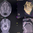
Using dental cone-beam computed tomography (CBCT) to measure bone density may serve as an effective method for screening osteoporosis in women, according to research presented at the International Association for Dental, Oral, and Craniofacial Research (IADR).
A clear link was found between the trabecular bone density of the right mandibular condyle (MCTBD) and the bone mineral density (BMD) of other joints during menopause, according to research presented at the 102nd General Session of the IADR, which was held in March in New Orleans.
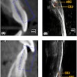
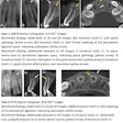
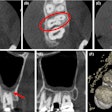
The investigation examined 357 deidentified CBCT DICOM files of dentate females in a cross-sectional study. They utilized ImageJ software 2.0 to analyze the gray values (GV) of the trabecular portion of bilateral mandibular condylar heads at their widest mediolateral dimension, according to the study led by Dr. Anh Le, PhD, of the University of Pennsylvania School of Dental Medicine.
Furthermore, the MCTBD was calculated using an internal calibration formula incorporating values from condyles, soft tissue, and air in the jaw. A paired t-test compared MCTBD values between the right and left sides. In a subset of 110 individuals who had both CBCT scans and dual-energy x-ray absorptiometry (DXA) scans within about five years, correlations were examined between MCTBD and hip/femur variables, according to the study.
Results showed a significant correlation (p-value < 0.001) between the MCTBD on the right side and total hip BMD, total hip T score, and femur T score. Patients with low bone mass had a 30% reduction in right MCTBD compared to healthy individuals, with no such difference observed on the left side. Between the ages of 45 and 60, a significant difference in MCTBD was noted between the two sides, with the right side exhibiting higher density, according to the study.
Additionally, a linear regression model involving right MCTBD, body mass index, and age demonstrated good predictive ability for low bone mass, with 80% sensitivity, 66.6% specificity, and 79% area under the receiver operating characteristic (ROC) curve, further supporting the correlating between CBCT scans and degenerative joint diseases.


