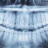
Periapical radiography is superior to cone-beam CT for detecting peri-implant bone defects, according to a study in Clinical Oral Implants Research (March 27, 2012)
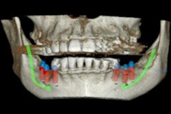
The researchers from King's College London Dental Institute and Guy's and St Thomas' Hospitals Foundation Trust placed implants in fresh bovine ribs in osteotomy sites of varying diameters: five with no peri-implant space; five with a 0.35 mm space; and five with a 0.675 mm space. They then imaged the sites using digital long-cone periapical radiographs, limited volume cone-beam CT (3D Accuitomo 80, J. Morita), and large-volume cone-beam CT (i-CAT Next Generation, Imaging Sciences International).
Images from each were randomly presented to nine examiners on two occasions. Confidence in diagnosing the presence or absence of a peri-implant radiolucency was recorded on a five-point scale.
The researchers found that the periapical radiographs were better at diagnosing a peri-implant bone defect when the peri-implant space was 0.35 mm (p < 0.02). As the peri-implant space increased to 0.675 mm, there was no significant difference in diagnostic accuracy between the three imaging methods. In addition, periapical radiography and the i-CAT had significantly better specificity and positive predictive value than the Accuitomo.
"Within the limitations of this study, long cone periapical radiographs are a reliable and valid method of detecting circumferential peri-implant bone defects and performed significantly better than [cone-beam CT]," the researchers concluded.