A growing body of research is lending weight to developers' claims that optical coherence tomography (OCT) can detect dental decay and other oral lesions better than traditional imaging modalities.
Researchers worldwide are now digging deeper to see what other dental applications OCT might be useful for. And the first commercial OCT systems for dentistry are moving closer to commercialization, with at least one product expected to launch in the first half of 2010, according to the manufacturer.
In the past 12 months, OCT research has expanded beyond validation studies that demonstrate the technology's ability to detect caries earlier than x-rays and spot fractures with greater precision. For example:
Researchers from the University of Kent collaborated with Romanian colleagues from Victor Babes University of Medicine and Pharmacy of Timisoara to evaluate the potential of OCT as an imaging tool for finding material defects in dental prostheses and microleakage at prosthetic interfaces (Journal of Biomedical Optics, September/October 2008, Vol. 13:5, 054065). Teeth were also imaged after several treatment methods to assess material defects and microleakage and to evaluate the quality of bracket bonding on hard tissue. Among their findings: Gaps between the dental interfaces and material defects were "clearly exposed."
A team from the department of orthodontics at Ulsan University Hospital in South Korea used OCT to study tooth movement under orthodontic forces (American Journal of Orthodontics and Dentofacial Orthopedics, February 2009, Vol. 135:2, pp. 252-259). Orthodontic distraction forces were applied to the mandibular incisors of six white rats for five days using individualized loop springs, and the periodontal ligaments were imaged with OCT and intraoral radiography. The researchers found that OCT allowed them to measure changed ligaments from all directions, while the radiographs could not show the portions overlapped by teeth.
At the 2009 International Association for Dental Research meeting in Miami, a team from Shahid Beheshti University in Iran and Tokyo Medical and Dental University presented their findings from experiments with OCT for in vivo assessment of composite restorations. The researchers used the swept-source OCT-2000 system from Santec to image a class III composite restoration in a patient 14 years after treatment. Visual examination showed that the restoration had mildly discolored margins, but cross-sectional OCT images showed that while enamel margins were sealed, there were some defects within the composite material and a loss of enamel and dentin up to 1 mm deep.
Also working with the Santec OCT-2000 system, researchers from Japan presented the first in vivo OCT images of oral vascular anomalies (British Journal of Oral and Maxillofacial Surgery, April 23, 2009). They analyzed vascular lesions in two outpatients at the National Center for Geriatrics and Gerontology in Japan, comparing OCT images to histopathologies. "OCT images correlated well with the histopathological sections, and we consider the system to be a safe way of imaging the oral microstructure; the ultrahigh resolution of the images enables improved visualization and segmentation of oral mucosal disease," they concluded.
Technological nuances
Other studies are looking more closely at the nuances of this evolving technology, comparing the pros and cons of the various types of OCT approaches now available and the wavelengths of light they use. This work could pave the way for additional dental applications and the development of more targeted tools.
For example, a team of Brazilian scientists and clinicians used an 850-nm spectral domain OCT system and a 1,280-nm time domain system to generate images of dentin and pulp chamber in in vitro teeth and compare them to cone-beam CT images (Journal of Biomedical Optics, March/April 2009, Vol. 14:2, 024009). According to their findings, OCT "clearly demonstrates the capacity of quantitative assessment and penetration of the radiation on the pulp chamber and the remaining dentin" compared to cone-beam CT. They also found that 1,280-nm OCT penetrates twice as deeply into tissue as 850-nm OCT.
 |
|
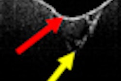
OCT captures cross-sectional images of hard and soft dental tissue up to 3 mm deep into the tissue. Image courtesy of Dr. Craig Gimbel, Lantis Laser. |
"OCT ... possesses great potential to be used routinely in clinical practice for the diagnosis of the complex dentin pulp, preventing accidental exposure of the pulp and promoting preventive restoration treatment," they concluded.
Meanwhile, researchers at the University of California, San Francisco have been studying the use of polarization-sensitive OCT (PS-OCT) to assess dentin demineralization and remineralization. In a study published this month, Daniel Fried, Ph.D., a professor of biomaterials and bioengineering in the department of preventive and restorative dentistry, and colleagues looked at the potential of PS-OCT to nondestructively measure the depth and severity of artificial demineralization on exposed root surfaces and measure the degree of inhibition by topical fluoride (Dental Materials, June 2009, Vol. 25:6, pp. 721-728).
They found that PS-OCT was able to measure a significant increase in the reflectivity between lesion areas and sound root surfaces. In contrast to dentin, they reported, the cementum layer manifests minimal reflectivity in the PS-OCT images, allowing nondestructive measurement of the remaining cementum thickness.
"With PS-OCT, we collect images in both polarization states; the light comes in at one orientation (parallel), and we measure it parallel and perpendicular to that," Fried explained. "It's just like how polarized sunglasses work -- you are removing glare from the light that is reflected. In the case of a tooth, there is very strong reflectivity from the tooth surface so we deliver the light in the parallel polarization because we only want to collect the light scattered inside the tooth, not reflected from the surface layer. With conventional OCT, because of the refractive index mismatch at the tooth surface, you get very strong reflectance, which can wash out the surface of the tooth."
On the market in 2010?
With this much attention being paid to OCT by the dental research community, clearly this is an imaging technology that is likely to have an impact in dentistry in the not-too-distant future.
The key to making OCT a clinical reality for dentists, however, is the development of a low-cost, compact, reliable system, noted Stanley Baron, president of Lantis Laser, which is developing an OCT system for dentistry. That is where Lantis has been focusing its resources since joining forces last year with Axsun Technologies, which has developed a new kind of OCT engine. Lantis expects to file a 510(k) application with the U.S. FDA this year and begin marketing its OCT system in the first half of 2010, Baron added.
"Lantis' OCT utilizes a spectral domain swept laser source, which is similar to the research devices being used at universities throughout the world," said Craig Gimbel, D.D.S., clinical director for Lantis and former president of the Academy of Laser Dentistry. Lantis is also developing near-infrared (NIR) imaging as an adjunct to the OCT system, through an exclusive license with the University of California.
"NIR will image the coronal tooth just like an x-ray but without any ionizing radiation and with far better resolution," Dr. Gimbel said. "NIR imaging will be used as a 'survey' macrostructural tool, and OCT will be used to image the microstructure of the oral soft and hard tissue."
Copyright © 2009 DrBicuspid.com