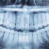
Could 3D ultrasound imaging improve the way periodontal disease is diagnosed and treated? A research team from West Virginia University (WVU) thinks so.
While ultrasound is best known in dentistry for its scaling and surgical abilities, the WVU team is studying the use of high-frequency ultrasound to construct 3D images of the mandible and surrounding tissues. They recently received a $393,575 grant from the National Institute of Dental and Craniofacial Research for a two-year project, Non-Invasive High-Resolution Diagnosis of Periodontal Attachment Levels Using Real-Time Quantitative Ultrasound Imaging.
Ultrasound imaging offers several advantages over conventional x-rays for periodontal applications, said Osama Mukdadi, Ph.D., an assistant professor of mechanical and aerospace engineering in the College of Engineering and Mineral Resources and lead researcher on the project. In particular, ultrasound can detect minute defects in bone that are difficult to see using radiographs, and it is safer because there is no exposure to ionizing radiation.
"X-rays have good resolution, but they are not sensitive to bone defects because of the shadowing, and they cannot provide enough information about periodontal disease at an early stage," Mukdadi told DrBicuspid.com. "Ultrasound has superior resolution, sensitivity, and contrast."
For example, at least 25% of the bone has to be gone before that loss can be seen on a radiograph, said Richard Crout, D.D.S., M.S., associate dean for research at the WVU School of Dentistry who is collaborating with Mukdadi and Peter Ngan, D.M.D., a professor and chair of orthodontics, on this project. So having a tool that can image early bone loss would be a boon for dental practitioners and patients alike.
"There is a pressing need for the development of better technologies for the early detection of periodontal disease," Dr. Crout said. "This technology provides more detail of the underlying bone surrounding the teeth than an x-ray but is less invasive since there is no radiation." Ultrasound imaging can also help determine what type of defect the practitioner is looking at, he added.
It also has some advantages over optical imaging techniques such as optical coherence tomography for periodontal applications, Mukdadi noted. "Optical imaging is a good solution, but it cannot see beneath the gingiva. We are interested in finding opaque objects or opaque defects, and optics doesn't help with that."
How it works
Ultrasound works by transmitting high-frequency sound pulses into tissue. As the sound waves travel, they hit a boundary between tissues (for example, between fluid and soft tissue or soft tissue and bone). Some waves are reflected back to the probe, while some travel on further until they reach another boundary and get reflected. The reflected waves are picked up by a probe and relayed to the machine, which calculates the distance from the probe to the tissue using the speed of sound in tissue and the time of each echo's return. The system then displays the distances and intensities of the echoes on the screen, forming a 2D image.
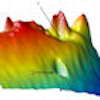
 Defected dentate dried cadaver mandible. Top, photographic image for the mandible with a rectangle showing the scanned region and three landmarks. Right, 3D ultrasound surface image for the jawbone surface for the rectangular region described in top image with landmarks. Images courtesy of Osama Mukdadi, West Virginia University College of Engineering and Mineral Resources. |
 |
The WVU ultrasound system goes a step further by providing 3D images. It employs high-frequency ultrasound-focused transducers (15 and 30 MHz) and a 1-GHz data acquisition card synchronized with a 2D stage positioning system. Signal processing algorithms are applied on the received ultrasound signals for filtering, focusing, and envelope detection prior to frame reconstruction, and an edge detection technique is used to detect the bone surface in each frame. The edges are combined to render a 3D surface image of the jawbone.
"The system utilizes a high-frequency ultrasound transducer that is capable of reconstructing 3D images of the jawbone," Mukdadi said. "These images reveal important information about opaque boney defects that could result due to periodontal diseases. The high frequency and thus high resolution images could detect tiny defects, which is important for early diagnosis of periodontal diseases."
Mukdadi's team has applied for a patent for this technology and is gearing up to begin clinical trials once their current research project is complete. A commercial version of the system could become available within two years at a cost of around $30,000 to $40,000, Mukdadi said.
Although the price might be off-putting to some, ultrasound's unique diagnostic capabilities would be an attractive addition to clinical dentists. Early detection can be a critical factor in the successful treatment of periodontal disease, said David Cochran, D.D.S., Ph.D., president of the American Academy of Periodontology and chair of the department of periodontics at the University of Texas Health Science Center at San Antonio, in a statement to DrBicuspid.com.
"It is always encouraging to see advances in technology, especially in imaging technology, that will allow periodontists to better serve our patients by catching the disease early and increasing their chances of achieving comprehensive oral health," he added.
Copyright © 2009 DrBicuspid.com