With its ability to create 3D images that contain key anatomical and morphological details, cone-beam CT (CBCT) is emerging as the standard of care for implant treatment planning and placement.
In fact, as more general dentists add implants to their list of treatment options, many are in turn investing in cone-beam CT systems and surgical guides to enhance implant outcomes, according to some experts.
— Scott Ganz, D.M.D.
"This is not a tool that should only be used by experts," said Scott Ganz, D.M.D., a prosthodontist in private practice in New Jersey who is also considered a leading expert on CT scan imaging and interactive 3D treatment planning. "By far it is a fabulous tool for people beginning to learn about implantology and endodontics because it teaches us that every patient's anatomy is different. Every patient presents with something different, and 2D technologies just don't give us enough information."
And a growing body of research indicates the dental community is embracing cone-beam CT in implant treatment planning for these very reasons. An article in the June 2010 Journal of the American Dental Association (Vol. 141:6, pp. 20S-24S), for example, concluded that "the precision of clinical treatment provided by the integration of CAD/CAM and CBCT allows dentists to plan for ideal surgical placement and the appropriate thickness of restorative modalities before placing implants."
Similarly, in the June 2010 Alpha Omegan (Vol. 103:2, pp. 51-56), an article entitled "3D cephalometrics: the new norm" concluded that "with the advent of cone-beam computed tomography (CBCT), the diagnostic foundations of dentistry have been forever changed and advanced into a new era of 3-dimensional (3D) possibilities. ... With CBCT imaging, the images are captured in their true anatomic size and shape, and therefore offer the most accurate treatment planning potential."
And a study in the August 2010 Implant Dentistry (Vol. 19:4, pp. 288-298) notes that "proper implant treatment planning remains the first priority for implant success. ... Traditional radiographs provide adequate information about proposed implant sites; however, limited film size, image distortion, magnification, and a 2D view restrict their use in some cases."
Multiple advantages
Cone-beam CT, on the other hand, provides important radiographic, restorative, and surgical information, including implant trajectory, distribution, depth, and proximity to critical anatomical landmarks, according to Allan Farman, B.D.S., M.B.A., Ph.D., D.Sc., a professor of radiology and imaging science at the University of Louisville in Kentucky and president of the American Academy of Oral and Maxillofacial Radiology (AAOMR).
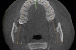
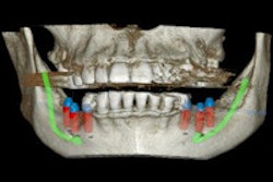
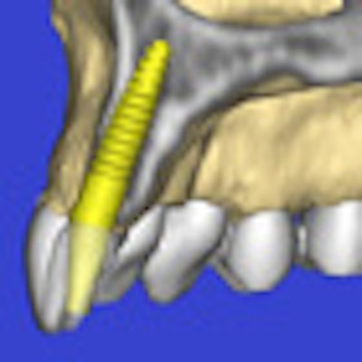
 Cone-beam CT provides important radiographic, restorative, and surgical information for implant treatment planning, including implant trajectory, distribution, depth, and proximity to critical anatomical landmarks. All images courtesy of Scott Ganz, D.M.D.
"One reason to do planning with 3D is to relate the implant to critical anatomical structures such as the mandibular canal, which contains nerve bundles that provide sensation to the lower lip," he said. "If impaired, a patient can have a loss of sensation in the lip, they can drool, and they will have reduced quality of life."
Using cone-beam CT for implant planning has nonclinical advantages as well, especially for those practitioners who are just becoming familiar with this procedure.
"There is plenty of research showing you can place implants and achieve a 90% success rate with panoramics or the other 'digital' ('two-finger) approach, especially if you've been placing implants for a long time," said Don Tyndall, D.D.S., M.S.P.H., Ph.D., director of oral and maxillofacial radiology at the University of North Carolina at Chapel Hill and head of the AAOMR committee that is revising the AAOMR position paper on imaging and implants. "But having the 3D information gives you a leapfrog of about 20 years' experience. It really helps give the beginner information that helps them with proper diagnosis and treatment planning, which gives them confidence. And in anything, if you have confidence, you usually are going to perform better and be less stressed."
Cone-beam CT also enhances patient acceptance, he added.
"I have heard so many times from cone-beam CT owners that the patient education aspect is huge," he said. "Acceptance rates increase significantly when the patient is able to see on a screen where the implant is going and the 3D space around it."
Surgical guides too
The growing adoption of cone-beam CT for implant planning is spurring a parallel increase in the use of surgical guides, Dr. Tyndall noted.
"Cone-beam CT sales are still largely driven by implant dentistry, although that is changing," he said. "But it is still the leading reason practitioners buy these systems. And in addition to the image data, they can generate surgical guides, which reduce surgery times and pain and increase healing times."
 |
| Click here to enlarge this image. |
| Integrated implant treatment planning systems such as the iCATVision can improve outcomes, encourage patient acceptance, and reduce treatment times. |
Cone-beam CT image data and surgical guides can also be integrated with in-office CAD/CAM systems such as the Cerec or E4D, Dr. Tyndall added, enabling dentists to diagnose, treatment plan, design, and fabricate all in the dental office. In fact, the vendor community has been one of the drivers behind the growing adoption of cone-beam CT in dentistry, Dr. Ganz noted.
"Implant companies have finally realized that if they embrace the technology, clinicians will learn how to assess the anatomy better and place implants better, then gain more confidence and place more implants," he said. "So the implant companies have finally developed their own product lines that take into consideration treatment planning."
It's not the scan that is the solution, "it's the plan," Dr. Ganz added. "It is the hardware that the implant companies are finally creating so you can accurately guide the drills and place the implants."
And like a growing number of his colleagues, Dr. Ganz sees cone-beam CT becoming the standard of care for implant planning, especially once it is taught in every dental school, every day, he said.
"This technology should NOT be used for only the difficult cases," he said. "It should be used for everybody, every case. Because it is the cases you think will give you no problems that often will give you the most problems, that surprise you because of the patient's anatomy. And you will be surprised at what you will see in a 3D cone-beam CT image."
Copyright © 2010 DrBicuspid.com