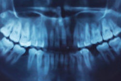
A majority of dental schools in the U.S. own cone-beam CT (CBCT) systems and have incorporated training in 3D image interpretation into their curriculums, according to research presented at the recent American Academy of Oral and Maxillofacial Radiology (AAOMR) meeting in San Diego.
With the growing interest in, and adoption of, cone-beam CT technology for maxillofacial imaging, researchers from Ohio State University and University of Detroit Mercy wanted to determine to what degree pre- and postdoctoral dental programs are including 3D image acquisition, interpretation, and application of implant planning software in their course offerings.
In April 2010, they e-mailed a nine-question survey to the instructors responsible for teaching oral and maxillofacial radiology at 57 dental schools and received a total of 56 responses.
"The oral and maxillofacial radiology academic community is a small group," lead author Vijay Parashar, BDS, DDS, MDSc, told DrBicuspid.com. "We all know each other almost personally, so we did not have a hard time getting the responses. We got most of them back within a month."
According to the survey results, 87% (50) of U.S. dental schools possess a cone-beam imaging system, and 91% include cone-beam CT in their predoctoral programs, according to Dr. Parashar, an assistant professor at Midwestern University College of Dental Medicine, who was originally at the University of Detroit when the project began. Six other schools said they are actively in the process of procuring a cone-beam CT system.
When asked if training in acquisition of cone-beam scans is provided to predoctoral students, only 18% responded yes. A large number of dental schools (82%) do not teach predoctoral dental students to acquire a scan or operate a cone-beam CT system. When asked if predoctoral students are educated in the interpretation of cone-beam CT scans, 48% responded yes and 52% responded no. Almost half of the schools teach cone-beam CT image interpretation to dental students to prepare them for identifying normal anatomy and abnormal pathology on 3D images.
Preoperative implant planning is one of the major indications for acquisition of CBCT scan in dental practices. Even so, only 32% of the schools teach predoctoral dental students to apply implant planning software, according to Dr. Parashar.
A higher number of schools (43%) teach to acquire cone-beam CT scans, and 81% of those providing postdoctoral dental education include training in cone-beam CT scan interpretation. In addition, 58% of dental residents receive training in application of implant planning software as part of their postdoctoral education.
"The majority of U.S. dental schools recognize the importance of teaching dental students about CBCT," the researchers concluded. "While most do not include instruction in higher level use of the technology for predoctoral students, larger numbers of schools provide this training to residents in specialty programs."
Dr. Parashar and his colleagues are now conducting a similar survey involving dental schools in the U.K. and Australia, working with researchers from King's College London and the University of Queensland, he noted.
They are also planning on sending a follow-up survey to the U.S. schools asking more specific questions about how many hours of cone-beam CT training each offers, what systems they use, whether they are small or large field-of-view, and other details related to the equipment and usage, Dr. Parashar said.
Copyright © 2010 DrBicuspid.com