Magnetic resonance imaging (MRI) is a promising alternative to cone-beam CT for locating the mandibular nerve prior to third-molar extraction, according to a study in Clinical Oral Implants Research (April 13, 2011).
The radiographic appearance of the mandibular nerve is radiolucent, and its course can be detected using the bony mandibular canal, which is radiopaque. The appearance of the canal is determined by the bone trabeculation density of the mandible. As a result, neither the mandibular canal nor the mandibular nerve are well-visualized in a radiograph when the mandibular trabecular bone is sparse, noted study author Anson Chau, BSc, MPhil, in the department of radiology at Prince Philip Dental Hospital in Hong Kong.
Both cone-beam CT and MRI have high accuracy in determining the location of the mandibular nerve, he noted. While MRI has the added advantage of not delivering ionizing radiation, so far it has not become a routine modality for dental imaging.
— Anson Chau, BSc, MPhil, Prince Philip
Dental Hospital
"Therefore, a comparison of the use of cone-beam CT and [MR] images in mandibular nerve detection among dentists who have lesser experience in imaging is needed because many disciplines now use cone-beam CT in their clinical practice," Chau wrote.
This study involved 11 patients who underwent cone-beam CT and MRI scans of the mandible prior to third-molar teeth extraction. Eight of the patients had horizontally impacted third molars. The cone-beam CT scans were taken using an iCAT system (Imaging Sciences International); the MRI scans were taken using a Magnetom Trio Tim syngo system (Siemens Medical Solutions).
Chau recruited 12 dental postgraduates in five disciplines to evaluate and compare the reliability of the locations of the mandibular nerves identified in the images. Among the assessors, two had no experience in reading either cone-beam CT or MR images, while the others had one to six years of experience reading cone-beam CT scans and two had some experience (four and 10 years, respectively) reading MR images.
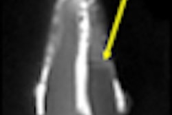
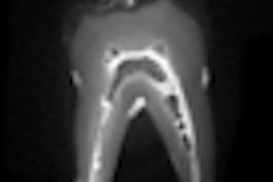
Each assessor identified the mandibular nerve, the mandibular foramen, or the mental foramen on 278 oblique cone-beam CT images and 298 oblique MR images.
Significantly lesser variances and significantly higher satisfactory scores were given to the MR images by the assessors than the cone-beam CT images. In six patients, significant differences were found in the mean of the variance of the x and y coordinates representing the locations of the mandibular nerve, mandibular foramen, and the mental foramen (p < 0.05). In addition, a significant difference was found in the satisfactory score between the use of cone-bean CT and MR images in identifying structures (p < 0.01).
"This study showed that [MR] images provide lesser variability in determining the locations of the mandibular nerve, the mental foramen, and the mandibular foramen than cone-beam CT images on patients who were scanned before wisdom teeth extraction," Chau wrote.
In addition, while the majority of assessors in the study had no experience in reading MR images, the interobserver reliability was still high, he noted. The assessors only used oblique cone-beam CT and MR images to identify mandibular nerve locations; adding multiplanar images and a reconstructed panoramic view could further enhance the identification of the mandibular nerve in clinical practice, he added, noting that image manipulation and processing techniques of MR images "were beyond the scope of this study."
"MRI provides high image contrast between the mandibular nerve and the bony dental structures," Chau wrote. "This decreased the variation in identifying the nerve location compared with the use of cone-beam CT images."
As a result, "MRI can be considered a promising alternative imaging modality applicable to many dental disciplines when the mandibular nerve of a patient cannot be identified on cone-beam CT images," he concluded.