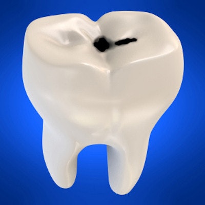
Near-infrared light transillumination (NILT) does not expose patients to ionizing radiation, but is it an effective modality for detecting caries? Researchers tested NILT against direct digital radiography in almost 140 teeth to find out.
NILT showed a higher correlation than direct digital radiography for approximal dentinal caries detection, and it can be a useful diagnostic tool in conjunction with bitewing radiographs, the study authors noted.
"Accordingly, NILT may be used to monitor the progression of caries without exposing the patient to ionizing radiation -- this being particularly interesting in growing patients and in pregnant women," wrote the authors, led by Maria Melo, DDS, PhD, of the Valencia University Medical and Dental School in Spain (Scientific Reports, October 2, 2019).
Caries detection
Near-infrared light transillumination is a photo-optical method for caries detection and diagnosis in posterior teeth. While the modality was first described in 1995, it did not reach the dental market until 2012. It uses visible light at a wavelength of 780 nm. In previous studies, the transillumination of tooth surfaces with light at specific wavelengths has shown good potential for detecting incipient lesions because it allows for differentiation between healthy and carious tissue. The researchers wanted to conduct an in vivo prospective study assessing NILT as a method for detecting approximal dentinal caries by comparing it with digital bitewing radiographs.
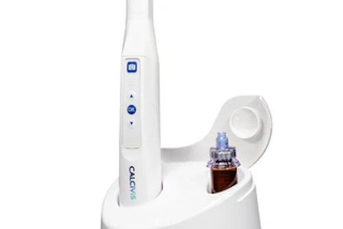
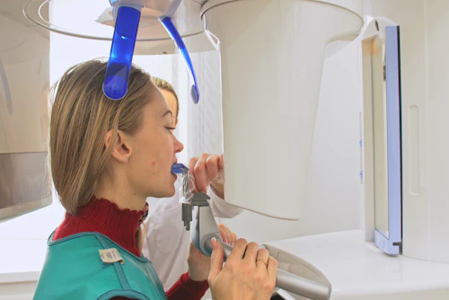
A prospective study was made of 112 patients at the University of Valencia Department of Dentistry in Spain. All the patients were seen for a routine dental visit and were evaluated with visual examination using the International Caries Detection and Assessment System (ICDAS), NILT (Diagnocam, KaVo Kerr), and the direct digital radiography system (Gendex Oralix, KaVo Kerr).
Researchers initially analyzed more than 1,280 teeth. The study included a total of 138 teeth (76 molars and 62 premolars) from 88 patients (49 females and 39 males).
The researchers found no significant difference in sensitivity between the two modalities. However, they did find a significant difference between them in correlation, with NILT showing a higher correlation (see table below).
| Differences among NILT, direct digital radiography, and ICDAS for sensitivity and correlation with histology | |||
| NILT | Direct digital radiography | ICDAS | |
| Overall sensitivity (n = 138) | 98.0% | 100% | 38.4% |
| Caries restricted to the outer half of dentin (n = 71) | 95.7% | 100% | 26.8% |
| Caries in the inner half of dentin (n = 67) | 100% | 100% | 50.7% |
| Correlation | 0.92 | 0.42 | 0.24 |
Clinical recommendation
The study authors reported they did not validate caries detected in enamel with direct digital radiography or NILT for ethical reasons. They listed no other study limitations.
The authors noted that none of the methods analyzed in this study can diagnose 100% of all carious lesions. The strength of direct digital radiography is precisely the main limitation of NILT: Direct digital radiography affords very precise visualization of the pulp chamber, but in the case of NILT, carious lesions at this level cannot be reliably diagnosed, they stated.
The researchers concluded by outlining their recommended clinical protocol.
"As clinical protocol, we recommend the ICDAS visual method to be used first (in the awareness that this only allows us to detect 38.4% of the lesions), followed by NILT. Only if NILT effectively diagnoses approximal caries do we then apply [direct digital radiography], thereby significantly reducing patient exposure to ionizing radiation," they wrote.