A cone-beam computed tomography (CBCT) scan helped identify a fifth canal in the first premolar of a man who was undergoing a root canal. This is the first reported case of a fifth canal in this type of tooth, according to a case report published on September 10 in BMC Oral Health.
The case highlights the need for clinicians to have a clear understanding of all tooth root canals to improve treatment outcomes. Also, CBCT should be used when a radiograph fails to show a clear image of the lower one-third of a tooth canal, the authors wrote.
"This is the first case presentation of a fifth canal of the mandibular first premolar and advances our understanding of variations in the anatomy of the mandibular first premolar," wrote the group, led by Ming Zhang, PhD, of Fujian Medical University in Fuzhou, China.
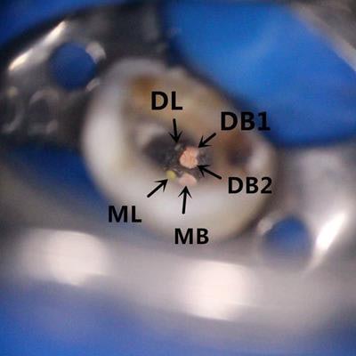
 Orifices of the five root canals of the tooth with obturation under the microscope. Mesiobuccal (MB), distobuccal 1 (DB1), distobuccal 2 (DB2), mesiodistal (ML), and distolingual (DL). All images courtesy of Ming Zhang, PhD, et al. Licensed under CC BY-NC 4.0.
Highly unusual
Branch root canals are common structure variations in root canal systems, presenting as one or more small lateral branches from the main root canal or as two equally large root tips that are divided into two forks on the apical segment. Clinicians have known about the anatomical variations in root canals of the mandibular first premolar. For example, in a study in an Egyptian population, approximately 97% of root canals in first premolars presented as a single canal; the rest had two canals. Other research showed that about 98% of mandibular first premolars had a single canal, 1.8% had two, and 0.2% had three. Less than 0.1% were found to have more than three canals.
A 25-year-old man
In the current case, an otherwise healthy man went to the hospital in April 2016 because he was experiencing intermittent pain in his lower right posterior tooth for more than three weeks. An exam and x-ray showed he had deep distant proximal and occlusal caries in the pulp chamber of tooth #44. This tooth showed slight knocking pain and sensitivity. There was no swelling, and the radiograph showed no apical radiolucency. He was diagnosed with symptomatic pulpitis and agreed to root canal treatment, according to the authors.
The man was placed under general anesthesia, and clinicians removed his caries and exposed the pulp chamber. The tooth was prepared for a root canal, and two weeks later the man returned to complete his treatment. Four root canals in the tooth were filled and sealed. During this process, a fifth canal located on the buccal side was found. A CBCT scan confirmed the fifth canal to be the mesiolingual root canal and curved. The fifth canal was sealed and filled, and x-ray and dental microscope images confirmed that the patient's canals had been fully filled.
A week later, the man was no longer experiencing pain. After two years, the tooth shows no signs of secondary caries, fractures, and apical radiolucency, the authors wrote.
 CBCT 3D reconstruction of the root canal of the tooth after it was filled. (A-D) 3D reconstruction of the tooth from different angles. The gray represents tooth tissue, and the red represents root canal tissue. (E) A cross-section of the root canal at the apical area.
Improving treatment
Root canal treatment for the first premolar can have a failure rate of up to about 11.5%. Therefore, successful treatment hinges on having a clear view of the canals of a patient's tooth. A CBCT scan can confirm the position of all canals in a timely manner, the authors wrote.
"This case advances the understanding of the variation in the anatomical structure of the mandibular first premolar and provides a reference for other similar cases," they concluded.