Editor's note: Allan Farman's column, Talking Pictures, appears regularly on the DrBicuspid.com advice and opinion page, Second Opinion.
The following message is typical of the type of inquiry I frequently receive. It has been adjusted to hide the identity of the person and place from which the inquiry was received -- and also the cone-beam CT (CBCT) vendor mentioned.
I am a general dentist in XXX, USA. Our office has a CBCT system, and we routinely take scans for implant patients and panographs. I was wondering if the CBCT can be used to detect caries by looking at small AP slices. We routinely do a FMX on new patients and then every five years. We take BWX every 12 to 18 months. I thought maybe, if caries can be detected accurately with CT, we could eliminate the FMX and do a CT and BWX. What is the difference in radiation exposure? Would this be a beneficial approach to eliminate the FMX?
Taking a full-mouth dental x-ray series as a baseline on new patients is acceptable so long as the patient does not have a recently exposed set of diagnostic-quality images taken elsewhere that can be utilized. Periodic bitewing radiographs at 12 months to 36 months to assess dental caries risk is also recommended by current FDA/ADA guidelines for prescribing radiographs for asymptomatic dental patients. All radiographs made should be based upon dental history, clinical inspection, and symptomology for the individual patient rather than as a routine.
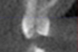
However, using cone-beam CT for dental caries detection is generally not recommended due to artifact from metallic and ceramic restorations making determination of disease, or lack thereof, precarious, to say the least. Because high-risk adult patients are likely to have such restorations, cone-beam CT would work best for those individuals at least risk. It is also unlikely to add information to the bitewing series.
Now, should cone-beam CT replace the full-mouth radiographic series or standard panoramic radiograph plus bitewings? In my view, we have not reached that point with current cone-beam CT systems and software. As noted above, cone-beam CT is generally not a good technique for dental caries detection, especially when teeth have been restored.
What about perio disease?
The second important condition for which dentists perform radiography is chronic periodontal disease. While cone-beam CT can demonstrate the periodontal bone in 3D, scatter artifact is still a problem when restorations are present. In addition, at lower resolution settings used to minimize radiation dose, voxel averaging can make precise interpretation of the presence or absence of thin anterior labial cortical plates difficult to ascertain.
Actually, the importance of cone-beam CT to periodontal inspection has been a cause of a recent discussion on the International Association of Dento-Maxillo-Facial Radiology Web site (specifically in the Oradlist group). While it is being used for this purpose in South America and some very nice images were presented on Oradlist, it was noted that referrals from U.S. periodontists for cone-beam CT services are almost exclusively for dental implant planning. It was also noted that there is presently no strong evidence that 3D imaging affects the outcome of periodontal disease treatment.
Based on these observations, I am inclined not to recommend cone-beam CT as a replacement for the periodic full-mouth radiographic series at this time. I regard cone-beam CT as an adjunct to, rather than a replacement for, traditional dental imaging methods.
Conditions in which I have found cone-beam CT useful include just about every case of dental implant placement planning; evaluating impacted third mandibular molars where the mandibular canal is superimposed on the roots of, or closely related to, the third molar on standard panoramic imaging; for certain endodontology purposes (with high-resolution scans) if traditional methods leave questions; and for delineating pathologic conditions affecting bony architecture.
For practitioners dealing with these conditions, access to a cone-beam CT system is undoubtedly of value. The question of whether to refer to a diagnostic center or own the system is a business decision predicated by a careful appraisal of utility. Given the costs of such systems, I would expect the practitioner making the purchase decision would to do so in an informed manner. Questions of whether dental caries can be detected should perhaps precede signing the check and passing it to the cone-beam CT vendor.
And radiation dose?
Regarding the question of radiation dose, the answer would depend upon the type of cone-beam CT system purchased. While many systems provide an order of magnitude less exposure to the patient than helical CT employed in most medical centers, this is not always the case. Dose is also dependent on the selected field-of-view (FOV) and the number of basis images selected. While the dose can be equal to or even less than a traditional panoramic exposure for small FOV systems, it would take the combination of up to six such small FOV images to provide a full panorama of the teeth, or three to six times the dose needed for the standard panoramic, to say nothing of the time to stitch adjacent volumes and examine the full dataset.
While making a cone-beam CT image takes relatively little time -- much less than exposing and processing a full-mouth intraoral series -- the exposure of the image dataset is but the beginning of the process. That dataset can certainly be processed within minutes. But then comes the construction of presentation states for evaluating the information that is present.
In one day last week, my evaluations of maxillofacial cone-beam CT volumes revealed cases of atherosclerosis causing stenosis of the internal carotid artery within the cavernous sinus, chronic mastoiditis with adjacent otosclerosis, osteoporotic degeneration of the cervical spine, severe temporomandibular joint osteoarthritis, tonsilloliths, pineal gland calcification, carotid atherosclerosis at the bifurcation of the internal and external carotid arteries, severe cervical osteoarthritis, cranial base discrepancy with the maxilla, and an aggressive multilocular benign neoplasm. In some of these cases, the findings led to referrals for treatment of likely importance to the general health of the referred patients. As an oral and maxillofacial radiologist, this was all in a day's work; however, I might well question the time availability for a general dental practitioner to seek and interpret most of these entities, as well as whether they even learned about them in dental school.
Cone-beam CT is a valuable adjunct for dental diagnosis. But it is an adjunct to, not a substitute for, traditional methods. Furthermore, thorough evaluation of the image volume needs both time and expertise. Racing cars are beautiful instruments with the professional behind the wheel in Indianapolis or Daytona, but not so good if the rookie driver were to take one out on the local interstate.
The comments and observations expressed herein do not necessarily reflect the opinions of DrBicuspid.com, nor should they be construed as an endorsement or admonishment of any particular idea, vendor, or organization.
Copyright © 2009 DrBicuspid.com
