An alignment device created by U.K. researchers may help practitioners better position each patient undergoing cone-beam CT (CBCT) imaging, eliminating the need for preliminary scout views prior to scanning and reducing radiation dose, according to the investigators.
In a paper published in Dentomaxillofacial Radiology (September 2010, Vol. 39:6, pp. 375-382), Veronique Sauret-Jackson, Ph.D., M.B.A., of Cavendish Imaging and three colleagues demonstrated that using the alignment device yielded positioning of the patients that was within a few millimeters of positioning produced by scout views.
"The new aligner will help position the patient's area of interest within the scanner's field-of-view (FOV) for situations where referrers require either large or very small imaging volumes, without the need for scout images," Sauret-Jackson told DrBicuspid.com. "Not only will this minimize radiation dose, but it will also help the endodontist or implant surgeon to avoid imaging structures that they may not necessarily have the training or experience to interpret."
 |
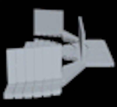
| The alignment device for positioning patients undergoing cone-beam CT imaging. Image courtesy of Veronique Sauret-Jackson, Ph.D., M.B.A. |
But Allan Farman, B.D.S., M.B.A., Ph.D., D.Sc., is skeptical about the practicality of the device. Dr. Farman is president of the American Academy of Oral and Maxillofacial Radiology (AAOMR) and a professor of radiology and imaging science at the University of Louisville School of Dentistry in Kentucky.
The investigators appear to believe that "smaller is better" when it comes to field-of-view, "rather than emphasizing that the appropriate FOV for each region of interest should be applied, whether large or small," he said. "And also, should volumes be collimated too greatly, there is always the possibility of needing additional doses while making retakes when one finds the original image will not suffice."
In addition, many of the cone-beam CT systems currently on the market cannot be tightly collimated, so this device would not work with them, Dr. Farman noted. He also expressed concern that infection-control issues could arise from placing the device in patients' mouths.
Dr. Farman noted that the AAOMR, together with other organizations such as the American Association of Endodontists and the American Association of Orthodontists, is producing guidelines on selection criteria for cone-beam CT images and image parameters. He believes these guidelines will be more effective in minimizing radiation dose than the use of a positioning device.
Sauret-Jackson responded that most cone-beam CT manufacturers now offer small- or medium-sized FOV designs, and that her team's new aligner will work with these.
"Those who have used the device -- which is, of course, autoclavable -- have highlighted the educational benefit," she said. "By making the cylindrical field-of-view more 'tangible' to the operator, the geometry and operation of the apparatus comes alive, improving understanding and hopefully also improving results."
Clinical test results
Sauret-Jackson and her team used computer-aided design software to create the specifications for the device. Then a series of prototypes was made by a small-run, rapid-manufacturing company in London.
The device has lines on targets on the bottom; using these, the radiographer can decide which line to align with the cone-beam CT apparatus' laser illuminators to target the center of the tooth or area of interest.
The team validated that the aligner device is accurate by placing the prototype in a plastic test object that they then placed in an iCAT scanner (Imaging Sciences International). Use of the aligner led to pinpointing the central axis of the FOV within 7 mm on the right-left and anterior-posterior directions and within 5 mm in the up-down direction, compared to using the scanner's laser illuminators.
Next, Sauret-Jackson and her colleagues recorded the accuracy of the aligner device and the laser illuminators of the Accuitomo F170 (J. Morita) in 47 patients. They first documented the set of coordinates from three directions using the scanner's scout views, then recorded the coordinates using the alignment device. Their statistical analyses revealed that the two methods produced similar measurements.
The second in vivo investigation involved two patients who were referred for visualization of a 5-cm-diameter area on the left upper first molar. The team used the iCAT scanner, which only projects the side illuminator onto the right-hand side of the patient's face. They then successfully used the aligner device to position the patient so that the region of interest was on the axis of the scanner's center of rotation.
Copyright © 2010 DrBicuspid.com