When trying to locate microcracks on root dentin after root canal prep, is microcomputed tomography (micro-CT) or cone-beam CT (CBCT) a more effective imaging modality? Researchers scanned 30 molars to find out.
They wanted to see how the two imaging methods compared in finding microcracks on root dentin, while also evaluating the frequency of dentinal microcracks after root canal preparation using the ProTaper Universal system. Their study was published in Microscopy Research and Technique (July 16, 2019).
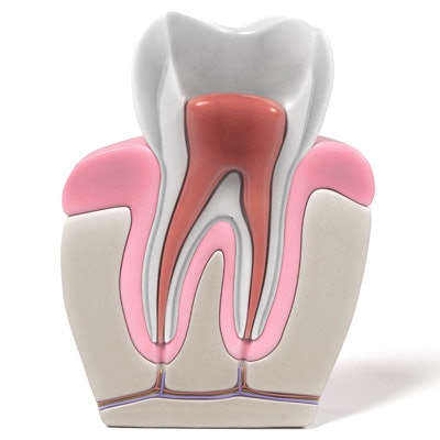
Microcracks that may form in dentin during some endodontic procedures can lead to vertical root fractures and may influence the long-term survival of endodontically treated teeth.
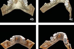
Researchers scanned the mesial roots of 30 extracted mandibular molars with both micro-CT and CBCT before instrumentation, which was then performed up to F2 and F4 ProTaper Universal files. They horizontally sectioned the roots at 2 mm, 4 mm, 6 mm, 8 mm, and 10 mm from the specimen apices and scanned again, then imaged the sections under a scanning electron microscopy for comparison.
The instrumentation process significantly increased the number of microcracks compared with preoperative samples, at least on micro-CT, which revealed that almost 44% of roots had microcracks. However, the scanning electron microscope revealed a significantly higher percentage of microcracks (88.3%), while no microcracks were observed using CBCT.
The table below shows the minimum, maximum, and median values of microcracks of pre- and postinstrumentation steps as found on micro-CT imaging. Instrumentation up to F2 and F4 file steps significantly increased the number of microcracks of the samples, the researchers noted. In addition, they found significant differences among preinstrumentation and first and second instrumentation steps.
| Percentage of microcracks found on micro-CT | |||
| Preinstrumentation | Postinstrumentation (up to 25 file steps) |
Postinstrumentation (up to 40 file steps) |
|
| Minimum (%) | 0% | 0% | 0% |
| Maximum (%) | 44.57% | 88.28% | 99.31% |
| Median (%) | 0% | 28.11% | 47.75% |
Many studies have evaluated vertical root fractures with CBCT, but this is the first study to assess if dentinal microcracks can be found with the modality, according to the study authors. Based on their study parameters, their research proves that dentinal microcracks cannot be detected accurately with CBCT.
They noted as a limitation, however, that it was an in vitro study.
The authors concluded that the right imaging method is essential to provide accurate results when searching for microcracks, and they noted that the preparation could lead to new microcracks.
"Root canal preparation up to F2 and F4 files with the [ProTaper Universal] system was found to induce the formation of new dentinal microcracks," wrote the study authors, led by Dr. İsmail Çapar, PhD, who is in private practice at the Periodent Dental Clinic in Istanbul, Turkey.