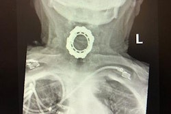
A 5 x 3-cm mandibular structure and eight teeth were found inside an asymptomatic ovarian dermoid cyst that was removed from a 15-year-old girl in New York, according to a case report published online on October 9 in the Journal of Pediatric and Adolescent Gynecology.
This is believed to be the first reported case of a cyst containing teeth within a mandibular-like bone in a pediatric patient. There are only five other cases like this in adults that have been reported, according to the authors.
"An adolescent girl found to have a mandibular structure with teeth in her dermoid cyst at the time of her laparoscopic ovarian cystectomy," wrote the authors, led by Dr. Stephanie Tardieu of the Icahn School of Medicine at Mount Sinai in New York City.
Dermoid cysts are congenital developmental anomalies and their etiology is mostly unknown. They are noncancerous tumors that occur when skin, hair, and other structures become trapped during fetal development. Though it is not unusual for an ovarian dermoid cyst to contain ectodermal derived tissue, including teeth, a cyst that presents with a fully formed mandibular structure with teeth is rare.
A 15-year-old girl
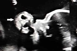
A girl visited the doctor's office for a consultation of a large asymptomatic right ovarian dermoid cyst that was detected on a sonogram when she went to the emergency room for a stomach virus. An ultrasound scan revealed a dermoid cyst measuring 10 x 8 cm in diameter with multiple echogenic areas, which doctors suspected were bone or teeth. The girl had no gynecologic or surgical history, she had no symptoms, and her physical exam was unremarkable. They agreed that a laparoscopic ovarian cystectomy would be performed, according to the case report.
At the time of surgery incisions were made and the cyst was visualized. They cut the right ovarian cortex and the cyst wall was identified. The cyst was removed and placed into a laparoscopic specimen retrieval bag, which was advanced to the left lower quadrant port incision site, they wrote.
When they cut into the cyst, they discovered the mandibular-like structure and eight teeth. They tried cutting and removing the mandibular-like bone with traditional gynecologic techniques using surgical scissors and a scalpel, but it posed the risk of damaging the instruments and failed to remove the specimen, the authors noted.
Creativity leads to a first
Clinicians had to figure out another way to extract the jaw-like bone and teeth, so they applied a novel technique to remove the cyst contents from the teen's abdominal cavity. They used an arthroscopic surgical blade to divide the mandibular-like bone into small pieces and remove them, which allowed for the procedure to be completed laparoscopically. Otherwise, the girl would have had to undergo a more invasive surgery to remove the specimen and likely experience more pain and require a hospital stay. This is the first known use of an arthroscopic surgical blade, which is used in orthopedic surgery to cut cartilage, joints, and bones, to debulk bony structures during a laparoscopic ovarian cystectomy, they wrote.
After the mandibular structure and cystic contents were removed from her stomach, the incision was closed. The patient was discharged from the hospital that day.
Lab results revealed that the cyst contained hair, fat, cartilage, and teeth, and the histological appearance of the jaw-like structure was mature bone, according to the report.
"Extracorporeal morcellation of bony structures within an ovarian teratoma using an arthroscopic surgical blade is technically feasible and led to successful completion," the authors wrote.