Clinicians removed a rare 4-cm mucocele that extended from the submandibular gland to the floor of the mouth of a 9-year-old girl. The case was published on June 16 in the Journal of Pediatric Surgery Case Reports.
This type of mucous-filled, cystlike lesion occurs in the oral cavity; it rarely occurs in the main salivary glands. This case is one of only 14 involving a mucocele in the submandibular gland reported during the past 30 or so years, the authors wrote.
"Mucocele of the submandibular gland is rare," wrote the group, led by Dr. Faiçal Slimani from Hassan II University of Casablanca in Morocco.
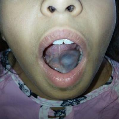
 The mucocele of the submandibular gland on the floor of the girl's mouth. All images courtesy of Slimani et al. Licensed under CC BY-NC 4.0.
Unique circumstances
The 9-year-old healthy girl had a swollen cystlike mass on the floor of her mouth and swelling in her left lower jaw area for about six months. She did not have any pain, and when the swelling began, she had no other symptoms.
Over time, the swelling increased in size but remained painless. However, the swelling started to have a mild effect on her speech, swallowing, and chewing, according to the authors.
Because her symptoms persisted, she was taken to a maxillofacial surgery unit. Her oral hygiene was borderline fair with some tartar and plaque and two caries. An oral exam revealed swelling of her left submandibular region that was soft and fluctuating, and the floor of her mouth was bulging from the lingual frenulum, elevating her tongue.
Closer inspection revealed a blue ovoid lesion that was painless with no secretion. A computed tomography (CT) scan confirmed the cystic mass, which clinicians determined needed to be removed.
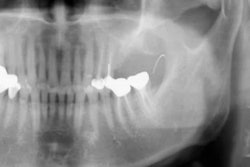
 Parenchymal window facial CT scan showing submandibular mucocele.
The child was placed under general anesthesia for removal of the cyst. First, her submandibular gland was removed, and then the entire mucocele was removed. The extracted 4-cm cystic mass was examined, and it corresponded histologically to a remnant salivary cyst. Lab results revealed that the submandibular gland was the site of "non-specific inflammatory fibrous changes," the authors wrote.
She was treated with antibiotics and discharged from the hospital after three days. Clinicians have followed her recovery for four years, and she has developed no complications or recurrence since her treatment.
Case importance
Typically, mucoceles are found on the inside of the cheek or lip or on the floor of the mouth. Though mucoceles usually are harmless, they can become uncomfortable over time, especially larger ones. Large mucoceles may also affect a person's speech, chewing, and swallowing abilities. If left untreated, they may cause permanent scar tissue.
Regarding a mucocele of the submandibular gland, "its topography, the site of other clinically similar conditions, and its clinical paucity require a rigorous clinical approach," Slimani and colleagues concluded.