A bone-encased dentigerous cyst was removed from a 6-month-old in New York, according to a case report published on July 22 in the International Journal of Oral and Maxillofacial Surgery. It is believed to be the first reported case involving an infant.
Dentigerous cysts are the most common developmental cysts occurring in the jaw over an unerupted tooth; however, they rarely occur before 10 years of age. Though panoramic x-rays can assist in diagnosing dentigerous cysts before patients develop symptoms and can prevent damage to other teeth, a change in pediatric x-ray guidelines likely isn't warranted, the authors wrote.
"Clinicians should be aware of the potential for this lesion to occur across a wide range of ages and the importance of prompt diagnosis and treatment to prevent complications and reduce morbidity," wrote the report's author, Dr. Monika Patel of the department of dental medicine at Northwell Health Long Island Jewish Medical Center in Queens, NY.
6-month-old boy with a bump
A 6-month-old boy with Factor X deficiency and a cleft lip that didn't require repair presented at the oral pathology department of a medical center in New York for a bump on his gums. The hematologist that the infant sees for his Factor X condition referred him to the center.
On examination, the cyst measured 2 cm and was overlying the lower left alveolus at unerupted teeth #74 and #75. The mass was bluish in color and extended from the lingual portion of the mandible laterally to the buccal vestibule. The overlying mucosa was intact, and the patient appeared to not be experiencing any pain.
Due to the Factor X deficiency, clinicians considered the lesion to be a possible vascular lesion and sent the infant to undergo magnetic resonance imaging (MRI). The MRI revealed a cystic lesion entirely encased in bone and surrounding the boy's permanent first molar tooth bud, according to the report.
The boy was administered general anesthesia and underwent a biopsy. Lab results of the cyst sample consisted of inflamed odontogenic cyst tissue linked with inflamed fibrous connective tissue and surgical hemorrhage. Parts of the tissue also exhibited reactive proliferative change with an associated underlying inflammatory infiltrate.
Based on the results, clinicians diagnosed the lesion as a dentigerous cyst and planned to remove the lesion from the adjacent bone, the authors wrote. Unfortunately, the boy was lost to follow-up between the time of diagnosis and treatment.
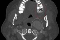
Nine months later, the boy was brought to the emergency department because he had acute left-sided swelling and intermittent intraoral bleeding, they wrote. He underwent a computed tomography scan that showed the cyst had expanded with a partially formed first permanent molar at the base of the cavity.
The patient was placed under general anesthesia, and clinicians enucleated the cyst, along with the permanent molar tooth bud, and extracted an adjacent baby tooth. Clinicians noted that the child's inferior alveolar nerve was intact under the cyst and was freed from the soft tissue, the authors wrote. Lab results of the entire lesion were like those seen in the biopsy sample except that it had more significant chronic inflammation, which is consistent with a dentigerous cyst.
Food for thought
Due to reported cases of dentigerous cysts arising into an ameloblastoma, an odontogenic keratocyst, and (very rarely) squamous cell carcinomas, it is critical that clinicians remove the entire cyst lining and submit it for a histopathologic exam. In most cases, dentigerous cysts increase in size, which can expand and weaken the surrounding bone and displace adjacent teeth, they wrote.
According to American Academy of Pediatric Dentistry guidelines, an x-ray should be taken after the first permanent tooth erupts. This may prevent the diagnosis of a pathology, such as a dentigerous cyst, until a patient has symptoms, like swelling, they wrote.
"Although an early panoramic radiograph at the time of completion of first molar crown development would have allowed timely treatment of this lesion, the rarity of such cysts at a young age makes it difficult to justify a change in pediatric guidelines for radiographs," Patel and colleagues wrote.