Being able to accurately describe oral mucosal lesions -- even when they're not seen often in daily practice -- can help dental practitioners better diagnose and manage them, and provide more comprehensive care for their patients.
— John McComb, B.D.S., M.Sc.,
head of oral pathology and oral
medicine, University of Toronto
That was the take-home message from three oral medicine experts at the recent Ontario Dental Association 2009 spring meeting in Toronto. During their three-hour session, "See, Describe, Manage: A Working Guide to Oral Medicine in General Practice," they dispensed easy-to-use tips for characterizing oral lesions designed to "replace lingering doubt with an active management strategy."
"In practice, lesions come out of the blue. And you can't run through everything that you learned maybe 10, 20, or 30 years ago in oral pathology and pick out the lesion; you have to use other principles to help you determine what it is," said John McComb, B.D.S., M.Sc., head of oral pathology and oral medicine at the University of Toronto. "Our goal isn't to teach you a long list of diseases. It's to give you a shorthand way to access clues to the identity of the lesions, the diseases of the mouth, that perhaps do not present in your practice every day."
Armed with this information, dentists should then be able to classify a lesion and decide whether it needs biopsy, referral, treatment, or observation.
The first task is to find a noun that describes the general form of the lesion. And the first question in that quest is whether it is a mass or an area of swelling. A mass is usually discrete and can be palpated, has a texture, and has limits, Dr. McComb said. Swelling, on the other hand, is more diffuse. Then, if it's a mass, determine the category. Is it a nodule, an ulcer, an erosion, or a crater? A macule, papule, path, or plaque? Or is it a small nodule, a vesicle, or a bulla?
Next, measure it accurately using a periodontal probe or ruler, and make a note of it. Also note its size and site, along with the distribution of the pathology if more than one lesion is present.
It is also important to describe whether the lesion is sessile or pedunculated; fixed or mobile; well-defined or diffuse; ovoid, circular, or has some other defined contour; what the texture or consistency is; and what its growth pattern is, Dr. McComb noted.
In fact, texture is often the most telling characteristic. "I tell my patients, 'If there's a thickening and a cardboardy type of texture (indicating a possible carcinoma), come and see me immediately,' " he said. "One of the things you need when you have a lesion under observation is help from your patient. Most sensible patients can be enormously helpful to us if they understand what we're trying to achieve."
Record the description
Karen Burgess, D.D.S., M.Sc., an assistant professor of oral pathology and oral medicine at the University of Toronto, said you should also write a description of the lesion while the patient is in the chair, so you can compare its appearance and size at the next visit or look it up in a textbook, such as Oral and Maxillofacial Pathology (3rd edition, W.B. Saunders, Philadelphia, PA, 2008).
According to Dr. Burgess, an example of a complete description is: "There was a blue, fluctuant swelling on the mucosal aspect of the lower lip opposite tooth 33. It measured 1.2 cm in diameter and appeared translucent. It extended into the superficial soft tissue of the mucosa." This is consistent with a mucous extravasation cyst, also known as a mucocoele, she noted. Additional signs, such as painless or pruritic, for example, may be uncovered during the history taking and examination, and incorporated into the description.
Other information that can help in assigning the lesion to a disease category include the following:
Duration: Lesions that are inflammatory, reactive, toxin- or drug-induced, malignant neoplasias, or, in some cases, immune-mediated, usually are present for only days or weeks before they become apparent to the patient or others; those that are cystic, benign neoplasias, hereditary, or developmental are often brought to a healthcare professional's attention only after months or years.
Presence of pain or tenderness
Rate of growth of a mass: Carcinomas, for example, may take months to reach a noticeable size, while sarcomas may double in size within just a few weeks. Focal fibrous hyperplasia, on the other hand, takes months or years to become visible/noticeable.
Presence of ulceration
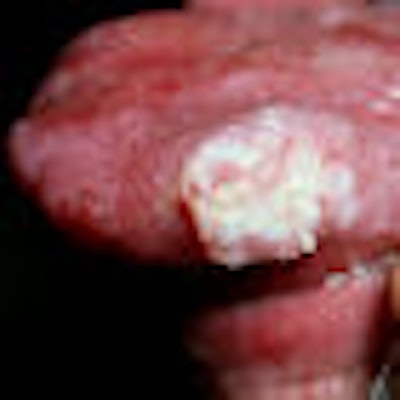
Dr. Burgess described a range of other keratotic and soft-tissue masses that can be found in the mouth, from pyogenic granulomas to squamous cell carcinoma. The latter is "the most serious lump or bump you can have in the mouth," she noted. The good news is that "they can be very successfully treated, especially if detected early," but they should be referred to a head and neck surgeon or an oral surgeon for treatment.
 |
| Squamous cell carcinoma of the tongue. Photo courtesy of Dr. Scott McComb. |
Iona Leong, B.D.S., an assistant professor of oral pathology and oral medicine at the University of Toronto, also provided a detailed description of pathologies that lie within four categories of ulcers: single acute, single chronic, multiple acute, and multiple chronic. The characteristics needed to determine the diagnosis include the following:
- Whether the lesion has been present for shorter or longer than three weeks (whether it is acute or persistent/chronic)
- Whether it is the result of a local influence, such as trauma from a jagged tooth surface or a systemic disease
- Whether one lesion or several are present; the anatomic location
- Whether it is shallow or deep
- What form the borders take; when they are rolled, for example, this may be a sign of a serious lesion such as a squamous cell carcinoma
- The patient's age
- Other etiological factors
Complaints of pain are "generally not helpful in the diagnosis of ulcers; in fact, malignant ulcers don't tend to present with pain unless they have invaded nerves or grown to a significant size," Dr. Leong said.
"A short list of general disease categories may cover more than 95% of the conditions you are ever likely to encounter," Dr. McComb concluded. "If you recognize something as belonging to the other 5%, then you are ready to refer -- and are probably well ahead of your peers in diagnostic acumen."
Copyright © 2009 DrBicuspid.com