A 27-year-old woman lost sensation in her chin and lip following root canal treatment on her mandibular molar. Months later, specialists confirmed that extrusion of the root canal sealer caused the patient's permanent anesthesia, according to a case report published April 1 in Oral Surgery.
Imaging showed that root canal sealer was widespread within the marrow spaces where endodontic treatment had occurred. The sealer extended to the mandibular canal and perforated the mandible's lingual cortex. There was also evidence of bony destruction and periosteal reaction.
"This case highlights the diligence that is required when performing root canal treatment, especially in cases where root apices have an intimate relationship with the mandibular canal," wrote the authors, led by Dr. Hanya Mahmood of the Academic Unit of Oral and Maxillofacial Surgery, School of Clinical Dentistry, at University of Sheffield in the U.K.
27-year-old woman with persistent numbness
The woman's numbness and tingling sensations began immediately after a general dentist performed endodontic treatment on tooth #47. About six weeks after the root canal treatment, the woman visited an oral surgeon, who extracted tooth #47 due to pain and infection.
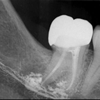
 On the left, a dental x-ray shows apical radiolucency, with superimposed radiopaque sealer material extruding at the root tips of endodontically treated tooth #47. On the right, the x-ray shows its presence in socket 47 after extraction. All images courtesy of Mahmood et al. Licensed CC BY 4.0.
However, pulling the tooth did not resolve the numbness. The patient reported recurrent biting and trauma to her lip, according to the authors. Periapical x-rays taken by the referring oral surgeon showed a radiopaque material at the root tips of tooth #47 and in the mandibular canal area after the tooth was pulled.
 Cone-beam CT shows the remnants of radiopaque sealer material left behind from previous endodontically treated and extracted tooth #47, with associated radiolucency suggestive of chronic infection.
The oral surgeon referred the woman to a trigeminal nerve injury clinic, which ordered neurosensory testing. The results revealed that the patient could not detect light touch or a sharp pinprick to her right lower lip and chin, indicating serious loss of sensation, according to the report.
A cone-beam computed tomography (CBCT) scan confirmed widespread extrusion of the root canal sealer in this region. Upon surgical exploration and decompression of the woman's right inferior alveolar nerve, clinicians found an avascular and fibrotic nerve with virtually no normal neuronal structure. They also saw evidence of foreign material, which was found to be root canal sealer, in contact with the epineurium and within the nerve trunk.
 The left image shows widespread fibrosis and necrotic nerve tissue in the apical region of extracted tooth #47. The right image shows foreign material on the external surface and within the nerve epineurium.
On a follow-up visit, the patient reported no improvement in her anesthesia symptoms. Since clinicians could not remove much of the endodontic sealer, the woman's nerve is not expected to recover, the authors wrote.
Preventing similar cases
Though damage to the inferior alveolar nerve due to endodontic treatment is rare, it can cause patients to permanently lose sensation in their teeth, chin, and lip. This outcome can be caused by overextension or overfilling of roots, the authors noted. Imaging is necessary to identify high-risk cases, provide accurate canal measurements, and assess the quality of root fillings, the authors noted.
The type of root canal filling system or sealer cement, the technique used, and nerve trajectory can all affect the incidence of this complication, the authors wrote. Previous evidence has shown that material extrusion may occur more often with bioceramic sealers than with those made of resin. With the potential rise in use of bioceramic sealers, which increase sealer volume, the risk of material extrusion may be greater, according to the report.
The case underscores the diligence required when performing root canals, specifically in patients who have teeth with root apices that are closely situated with the mandibular canal, Mahmood et al wrote. If endodontic material extrusion occurs, practitioners should urgently refer patients to an oral surgeon for surgical decompression to maximize the potential for nerve regeneration.
"Whilst the prevalence of [inferior alveolar nerve] injuries secondary to endodontics is low, with only a limited number of published case reports, it is important for clinicians to be able to recognize which teeth pose a greater risk and how to expedite management should such a complication arise," Mahmood and colleagues concluded.