Imaging helped treat a healthy 17-year-old girl's pseudoaneurysm, a rare, life-threatening vascular lesion, that developed following routine third-molar extractions. The case report was published in the March issue of Oral and Maxillofacial Surgery Cases.
A few days after the extractions, the teen developed mouth pain and uncontrollable bleeding. A computed tomography angiography (CTA) showed a lesion emerging from the girl's inferior alveolar branch of her right internal maxillary artery. An image-guided procedure was used to treat her, the report's authors wrote.
"Any practitioner performing dental extractions who encounters unusual bleeding should be aware of this potentially fatal vascular lesion," wrote the authors, led by Dr. Jiean Heifetz-Li, MPH, of Temple University Hospitals, Oral and Maxillofacial Surgery, in Philadelphia (Oral Maxillofac Surg Cases, March 2022, Vol. 8:1, e100249).
Teenager with uncontrollable bleeding
A 17-year-old girl with no significant past medical history went to an oral and maxillofacial surgery clinic to have her impacted wisdom teeth extracted. Surgically removing her mandibular third molars was considered a routine procedure.
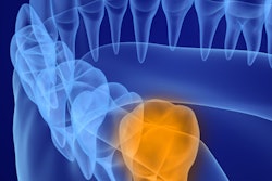
 Pre-extraction panoramic x-ray showing complete bony impacted third molars. All images courtesy of Heifetz-Li et al. Licensed by CC BY 4.0.
A distal "hockey stick" incision was used to raise the mucoperiosteal flap to get to the crowns, which were lingually inclined. The nerves were intact, and the teeth were removed with no excessive bleeding, according to the report.
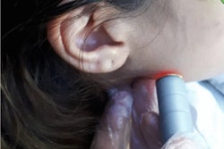
Three days later, the girl returned complaining of right facial swelling and bruising, pulsating pain, malaise, and fever. An intraoral exam revealed very inflamed soft tissue around the residual socket of extracted tooth #32 that was bleeding spontaneously and was tender to touch, they wrote.
Examining tooth #32's mucoperiosteal flap, the surgeon encountered brisk bleeding. Underneath the tooth's surrounding soft tissues were "liver clots," or incomplete fibrins clotting, which presented as slowly developing reddish-brown clots. No purulence was encountered, and the buccinator muscle was very bruised, the authors wrote.
Transfer to the hospital
After the tooth socket's base was cleared, clinicians determined that the girl's inferior alveolar artery was causing the bleeding. Unable to control the bleeding with oxidized regenerated cellulose, a sterilized compressed sponge, and an adjunct hemostatic agent, emergency medical services transferred her to an inpatient setting, they wrote.
Upon admission, the patient underwent a CTA with intravenous contrast of the maxillofacial complex. The imaging revealed a peripherally enhanced collection of fluid measuring 1.5 × 2.5 cm around the buccal and lingual surfaces of the mandibular body. Also, there was cortical erosion of the medial surface of the right mandibular ramus and vascular blushing of the inferior alveolar artery.
 A CTA maxillofacial with contrast shows a peripherally enhanced collection adjacent to the extraction socket of tooth #32, as well as cortical breakthrough along the medial surface of the right mandibular ramus.
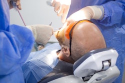
At this point, clinicians consulted with interventional imaging specialists who performed an angiography of her right external carotid artery under general anesthesia, according to the report.
The imaging showed contrast extravasation and a pseudoaneurysm emerging from her inferior alveolar branch of the right internal maxillary artery.
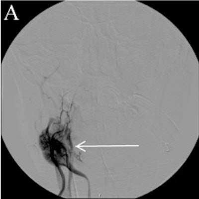
 A, B: An interventional cerebral angiogram shows contrast extravasation and a pseudoaneurysm arising from the inferior alveolar branch of the right internal maxillary artery (see arrow, panel A). C, D: Angiogram following coil embolization across the origin of the right inferior alveolar artery (see arrow, panel C and D).
The team placed a rapid transit microcatheter over a guidewire into the right maxillary artery, and a series of detachable coils were deployed across the inferior alveolar artery. After the procedure, the girl was transferred to the neurosurgical intensive care unit for continued observation.
On the second day after the procedure, the teen was given a blood transfusion in response to a decrease in her hemoglobin level from 12 g/dL to 9 g/dL. On the fourth day after surgery, the girl was discharged with close follow-up with oral and maxillofacial surgery and interventional radiology services, the authors wrote.
Common procedure, uncommon complication
In the U.S., oral and maxillofacial surgeons and general dentists extract more than 10 million impacted third molars each year. Complication rates for these procedures range from 3.5% to 14.8%. The most common postoperative complications for wisdom tooth extractions include infection, alveolar osteitis, and paresthesia.
Pseudoaneurysms, like in this case, are perivascular hematomas that often form after arterial injury and are retained by a fibrous pseudocapsule. Though they can be found anywhere in the body, most pseudoaneurysms are found in the femoral artery following catheterization and occur at an incidence rate of 0.6% to 6% of patients. Due to their higher rates of rupture, pseudoaneurysms are considered vascular emergencies and carry the potential to result in life-threatening hemorrhage.
In dentistry, most reported cases of pseudoaneurysms are diagnosed in patients with either sustained recent maxillofacial trauma or following microvascular or orthognathic surgery. Though significant postoperative bleeding is a rare complication following third-molar extractions, clinicians should be aware of the causes and potential treatments, the authors wrote.
In this patient case, the girl reported intermittent headaches in the months after the procedure that became less frequent over time. She has had no recurrent incidents of bleeding since its initial presentation and, fortunately, no problems with the reception and interpretation of sensory stimuli.
"While the most common cause of postoperative bleeding is soft tissue injury, pseudoaneurysms, although rare, should be included in the differential diagnosis when bleeding persists despite continued local measures," Heifetz-Li and colleagues concluded.