A healthy woman developed the extremely rare complication of traumatic myositis ossificans of the temporal muscle attachment after receiving an injection of local anesthesia near the right temporal muscle attachment for root canal treatment. The case report was published on June 22 in Clinical Case Reports.
Following the dental procedure, the woman experienced persistent limited mouth opening. Eventually, she made a full recovery after undergoing a coronectomy and engaging in physical therapy and regular jaw exercises, the authors wrote.
“The diagnosis could be considered for patients presenting with therapy-resistant trismus after intraoral procedures,” wrote the authors, led by Dr. Torbjørn Østvik Pedersen of the department of maxillofacial surgery at Haukeland University Hospital in Bergen, Norway (Clin Case Rep, June 22, 2023).
A healthy woman in her 30s
Prior to undergoing root canal treatment of her maxillary premolar, the woman was administered local anesthesia near her right temporal muscle attachment. Immediately following the injection, the woman reported experiencing a sharp pain and persistent limited mouth opening after treatment was completed, the authors wrote.
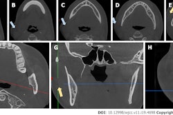
Four months after treatment, the woman presented with an inability to open her mouth. At that time, her mouth opening was 8 mm. The patient was unable to move her jaw laterally. She underwent a computed tomography (CT) scan, which showed an approximately 3-cm long bony growth that extended from the right coronoid process of the jaw to the cranial base, they wrote.
 CT in the (A) frontal and (B) sagittal view shows ossification of the woman's right temporal muscle attachment extending to the cranial base (arrows). Images courtesy of Pedersen et al. Licensed by CC BY 4.0.
She underwent a coronectomy to partially remove the bony lesion, and a sample was sent to the lab for analysis. The histological diagnosis of normal bone tissue, coupled with the imaging results from the CT scan, led to the clinicians’ diagnosis of traumatic myositis ossificans, the authors wrote.
Immediately after surgery, the patient had a mouth opening of 32 mm. After one month, her maximum mouth opening dropped to 14 mm, although no bony interferences were detected on imaging.
After physical therapy and regularly exercising her jaw, she had a mouth opening of 27 mm and acceptable function at a postoperative visit five months after the surgery, according to the report. At a five-year follow-up, the woman had a mouth opening of 40 mm and was chewing normally, the authors wrote.
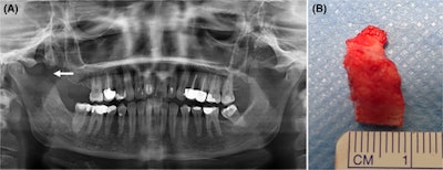
 The (A) panoramic x-ray after surgical treatment shows partial resection of the lesion and the coronoid process (arrow). A (B) clinical image of the resected lesion.
Highly unusual condition
Traumatic myositis ossificans is a rare condition that causes bone to form deep within a muscle. The condition is very unusual in the head and neck region. Most commonly, the masseter muscle is affected, and in most of the reported cases, it occurs following minor surgeries, like third-molar extractions.
When it does occur, typically the onset of symptoms begins three to six weeks after local muscle trauma. Additionally, there is limited research on the disease occurring following a dental procedure. However, there have been reports of traumatic myositis ossificans affecting the medial pterygoid muscle following mandibular nerve block, the authors wrote.
“This patient developed ossification of the temporal muscle attachment following infiltration of local anesthesia -- an extremely rare complication,” Pedersen and colleagues wrote.