Blind for nine years, Sharron "Kay" Thornton has regained her sight through a surgical procedure performed for the first time in the U.S. at Bascom Palmer Eye Institute at the University of Miami Miller School of Medicine.
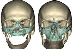
The procedure -- modified osteo-odonto-keratoprosthesis (MOOKP) -- implanted one of her canine teeth in her eye as a base to hold a prosthetic lens.
"I'm looking forward to seeing my seven youngest grandchildren for the first time," said Thornton, age 60, of Smithdale, MS, who was blinded by Stevens-Johnson syndrome in 2000, said in a press release. The rare skin condition destroys the cells on the eye surface, causing severe corneal scarring.
On Labor Day weekend, after the last in a series of surgeries by corneal specialist Victor Perez, M.D., an associate professor of ophthalmology at Bascom Palmer Eye Institute, bandages were removed from Thornton's eyes and she was able to recognize faces only hours after her surgery, according to the institute. Two weeks following her surgery, she is already reading newsprint with a visual acuity of 20/70, and her vision is expected to improve further as her surgical scars heal.
Developed in Italy, the procedure has proved effective as a solution to end-stage corneal disease in which severe corneal scarring blocks vision and corneal transplants are no longer an option, but the eye's internal structures and optic nerve remain healthy.
"For certain patients whose bodies reject a transplanted or artificial cornea, this procedure 'of last resort' implants the patient's tooth in the eye to anchor a prosthetic lens and restore vision," said Dr. Eduardo Alfonso, M.D., chairman of Bascom Palmer Eye Institute, in the release. "In Sharron's case, we implanted her canine tooth, her eyetooth."
 |
| Click here to enlarge this image. |
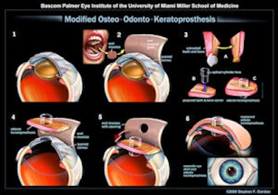
| In modified osteo-odonto-keratoprosthesis (MOOKP), a patient's canine tooth is implanted in the eye as a base to hold a prosthetic lens. Graphic courtesy of Business Wire. |
With the procedure, the tooth and surrounding bone are shaved and sculpted, and a hole is drilled for inserting an optical cylinder lens. Next, to bond the tooth and lens as a biointegrated unit, they are implanted under the patient's skin in the cheek or shoulder. Meanwhile, the ophthalmologist prepares the surface of the eye for implantation of the prosthesis by removing scar tissue surrounding the damaged cornea.
A month later, mucous material is collected from the inside of the patient's cheek and used to cover and rehabilitate the surface of the damaged eye. In the final phase, usually two months later, the prosthesis is removed from the cheek or shoulder and implanted in the eye. The prosthesis is carefully aligned with the center of the eye, and a hole is made in the mucosa for the prosthetic lens, which protrudes slightly from the eye and enables light to re-enter the eye, allowing the patient to see again.
In 2008, Dr. Perez traveled to Europe for training by Italian ophthalmologist Giancarlo Falcinelli, M.D., who developed the MOOKP procedure, building on the original OOKP technique developed in the 1960s by Benedetto Strampelli, M.D., also of Italy.
Copyright © 2009 DrBicuspid.com